Playlist
Show Playlist
Hide Playlist
Erosive Arthritis & Calcium Pyrophosphate Deposition Disease (CPPD)


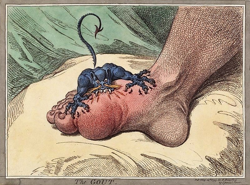
00:01 Erosive arthritis is characterized by inflammation and pannus formation. 00:05 Pannus is actually an enlarging synovial tissue which causes the erosions of the adjacent cartilage and bone and this actually includes many different types of arthritis. 00:15 The most common forms of erosive arthritis include rheumatoid arthritis, gout and psoriatic arthritis. 00:22 So rheumatoid arthritis is an inflammatory type of arthritis and it's most commonly seen in younger females. 00:29 The hands are the most commonly affected and usually sometimes the only affected area. 00:36 It can cause joint space narrowing with subchondral changes which are similar to that seen in osteoarthritis and usually the DIP joints of the hand or the distal interphalangeal joints are spared. 00:48 So as you can see in this patient, this is the metacarpal phalangeal joint. 00:53 It's the one that's most affected and then the PIP joint or the proximal interphalangeal joint can also be affected. It can cause periarticular osteoporosis which we can see in this patient here where we have the PIP joint and surrounding it, you have areas of lucency or osteoporosis. 01:12 It can result in soft tissue swelling and you can see erosions of the wrist and the proximal joints of the hand. 01:18 It can also cause ulnar deviation or subluxation at the MCP joints which we see in this patient here. 01:25 So this is the ulnar aspect of the hand and you can see that each of this MCP joints we have deviation towards the side of the ulna. 01:34 Gout is caused by deposition of calcium urate crystals within the joint space. 01:38 You can actually have many years of gout prior to the actual visualization of radiographic changes. This is most commonly seen in males and it most commonly affects the first metatarsal phalangeal joint or the MTP joint. 01:52 So here we have a coned down radiograph of the foot and it shows you the left MTP joint here. 02:01 You can see that there are erosive changes at the MTP joint which is characteristic of gout. 02:07 We have juxta-articular erosions and the classic term that's used is overhanging edges, so you have erosions that actually have edges that kinda hang over the joint space. 02:18 You can have tophi which are soft tissue collection of urate crystals and rarely this calcifies, so often they might not be seen in radiographs. 02:27 In the elbow, it can actually cause olecranon bursitis. 02:30 Psoriatic arthritis is often associated with skin and nail changes. 02:35 Typically, it involves the DIP joints of the hands. 02:38 So this is an example of a patient with psoriatic arthritis. 02:44 You can see here these are called pencil in cup deformities so you have narrowing of one of the bones while the other bone appears to form a cup around the area of narrowing and this is very characteristic of psoriatic arthritis. 02:57 You can see that it commonly affects the DIP joints or the distal joints of the hand. 03:01 It can also cause juxta-articular erosions and it causes enthesophytes or bony overgrowths at the site of tendon insertions. 03:09 Again, you can see this pencil in cup deformity which is the classic description of a patient with psoriatic arthritis and that's bony resorption of the terminal phalanges with one phalanx protruding into the other. 03:20 So CPPD or Calcium Pyrophosphate Deposition Disease is a mixed hypertrophic and erosive arthritis. 03:29 It's caused by depositon of calcium pyrophosphate dihydrate crystals within the cartilage surrounding the joints and it's often associated with chondrocalcinosis which is calcification of the articular cartilage. 03:41 Imaging features make it difficult to distinguish from osteoarthritis. 03:46 It does have chondrocalcinosis which we'll take a look at and the key feature is that it affects joints that are not typically affected by osteoarthritis, so similar radiongraphic findings as osteoarthritis but in joints, that you really wouldn't expect that to occur in. 04:00 You can also have pannus formation with CPPD. 04:03 So this is an example of chondrocalcinosis. 04:07 You have calcification of the articular cartilage which is pointed out by this arrow here. 04:11 This is commonly seen with arthritis in general and it's often asymptomatic but it is one of the features of CPPD. 04:19 So this is an example of a patient with CPPD. 04:22 In the hand and wrist, it has typical findings of what are called hook shaped overgrowths of the metacarpal heads and you have narrowing of the radiocarpal joint space with widening of the scapholunate junction. 04:33 We'll take a better look at these findings in just a second. 04:36 This can actually result in scapholunate advance collapse or SLAC wrist which we see here which is collapse of the capitate which is right here towards the radius. 04:46 So this is an example of the overhanging edges that you would see and here as we said we have collapse of the capitate towards the radius and you have narrowing of the radiocarpal joint space. 04:59 Here we have widening of the scapholunate junction. 05:02 So we have the lunate right here and we have the scaphoid here and this junction here is wider than you would normally expect.
About the Lecture
The lecture Erosive Arthritis & Calcium Pyrophosphate Deposition Disease (CPPD) by Hetal Verma, MD is from the course Musculoskeletal Radiology. It contains the following chapters:
- Erosive Arthritis
- Calcium Pyrophosphate Deposition Disease (CPPD)
Included Quiz Questions
What is TRUE about gout?
- It causes juxta-articular erosions with overhanging edges.
- It causes periarticular osteoporosis.
- It causes subchondral sclerosis.
- It causes osteophytes.
- It causes chondrocalcinosis.
Which of the following is NOT seen in erosive arthritis?
- Osteophytes
- Enlarged synovial tissue
- Pannus formation
- Cartilage erosion
- Inflammation
Which of the following is a typical feature of calcium pyrophosphate deposition disease (CPPD)?
- Chondrocalcinosis
- Pannus formation
- Subchondral cysts
- Widening of the joint space
- Erosive changes
Author of lecture Erosive Arthritis & Calcium Pyrophosphate Deposition Disease (CPPD)

Hetal Verma, MD
Customer reviews
5,0 of 5 stars
| 5 Stars |
|
1 |
| 4 Stars |
|
0 |
| 3 Stars |
|
0 |
| 2 Stars |
|
0 |
| 1 Star |
|
0 |
well explained, concise, not over complicated. I found it really engaging!
