Playlist
Show Playlist
Hide Playlist
Elbow Joint

-
Slide Elbow Joint.pdf
-
Download Lecture Overview
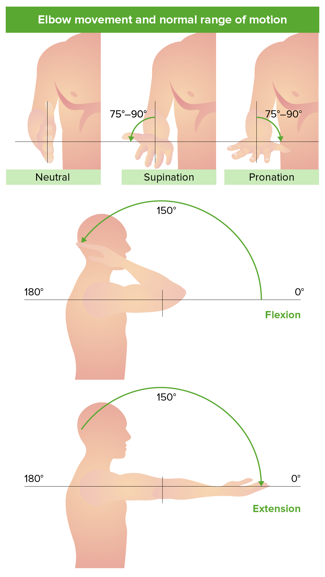
00:01 So, now, let's turn our attention to the elbow joint. 00:04 We've spoken a lot about the elbow joint and the movements of the elbow joints. 00:09 Now, let's have a look at it in some detail. So, here, we can see the three bones that form the elbow joints. 00:16 Here, we can see the distal aspects of the humerus and we can see the proximal aspects of the radius which is laterally and the ulna which is medially located. 00:27 So, here, we're looking at an anterior surface of the right elbow joint. 00:31 Here, we can see the surfaces for articulation between the trochlea of the humerus and the trochlear notch on the ulna. 00:39 And then, we can see how these two come together to form the humeroulnar joint. 00:44 Similarly, on the lateral aspect, we have the capitulum of the humerus and how that articulates with the head of the radius, forming the humeroradial joint. 00:54 So, although we're talking about the elbow joint, actually, you can see there's some very specific joints here, humeroulnar joint between the ulna and the humerus and the humeroradial joint between the radius and the humerus. 01:07 Here, we can see we have a nice joint capsule that is surrounding all of this joint and we can just make out the radial fossa which we can see there. 01:14 Remember, that allows the articulation, the rotation of the head of the radius during pronation and supination. 01:20 And here, we can see the coronoid fossa which allows the ulna to sit during full flexion of the forearm against the humerus. 01:28 It allows the ulna to position against that humerus there, the coronoid fossa. 01:33 Here, we can see the medial epicondyle and we can see the head of the radius, the coronoid process and all of these are completely surrounded by that joint capsule. 01:45 If we look more at the posterior aspect, you can still see the make out of the joint capsule there. 01:50 You can see the olecranon that's going to sit on the olecranon fossa during full extension of the elbow. 01:55 And here, again, you can see a number of ligaments similar to the glenohumeral joint that are here to support the elbow joint. So, in the medial aspect, you have a thickening of the joint capsule, the ulnar collateral ligament and we also have that on the lateral aspect with the radial collateral ligament. 02:13 These helping to support the joint capsule. 02:16 Running around the head of the radius, attaching to the ulna, and then, running all the way around in this C-shaped orientation, we have the annular ligament of the radius. That helps to hold the radial head in position. 02:28 Be careful when you pull the arms strongly of a young child because that ligament is still loose, so, you can pop the radial head out of that ligament and dislocate it. 02:37 So, here, we can see the movements of the elbow joint. 02:39 We're familiar with these already when we looked at the muscles of the arm. 02:43 We have extension of the elbow join in this direction and we have flexion of the elbow joint as well. 02:49 Blood supply, we mentioned these when we looked at the blood vessels of the upper limb. 02:54 But principally, we've got the brachial artery, the ulnar artery, and the radial artery, forming combinations of anastomotic pathways that supply this region. 03:05 So, here, we can see we have the brachial artery which is bifurcating down into the ulnar and the radial arteries and coming off it are a series of supporting branches. 03:14 So, superiorly, we can see the superior ulnar collateral artery that's also being supported by its sibling, the inferior ulnar collateral artery and we can see these are supplying the joint capsule. 03:27 And then, coming off the ulnar artery, we can see we have both a posterior and anterior ulnar recurrent artery. 03:35 And these are coming from the ulnar arteries and then, running superiorly up to supply the joint capsule. 03:41 On the lateral aspect, we have the radial artery. 03:44 We will return to the radial recurrent artery that runs back superiorly up to supply the joint capsule. 03:50 And here, we can also see now in the posterior surface, a number of branches which are more clearly coming to supply the more posterior aspect. 03:59 Coming off the brachial artery, we now see that superior ulnar collateral artery again and its posterior sibling. 04:05 We can now see a medial collateral artery coming off and we can see the radial collateral artery as well, this time, supplying the joint capsule we can see on this posterior aspect. 04:17 So, a whole series of very small thin branches coming off the key brachial, ulnar, and radial arteries of the upper limb. And these very importantly form an anastomotic network around the joint capsule, ensuring that joint capsule receives plentiful blood supply.
About the Lecture
The lecture Elbow Joint by James Pickering, PhD is from the course Joints of the Upper Limbs.
Included Quiz Questions
Which statement concerning the elbow joint is correct?
- The radial/lateral collateral ligament is continuous with the annular ligament.
- The capitulum articulates with the notch of the trochlea.
- The coronoid fossa limits hyperextension.
- The carrying angle in women is approximately 25 degrees.
- Its bony components are the humerus and the radius.
Which artery does NOT participate in the anastomotic blood supply network to the elbow joint?
- Axillary artery
- Radial artery
- Ulnar artery
- Brachial artery
What structure lies on the posterior part of the elbow?
- Olecranon
- Coronoid process
- Medial epicondyle
- Head of the radius
- Radial fossa
Which part of the humerus participates in the humeroradial joint?
- Capitulum
- Head
- Trochlea
- Trochlear notch
- Olecranon
Author of lecture Elbow Joint

James Pickering, PhD
Customer reviews
5,0 of 5 stars
| 5 Stars |
|
5 |
| 4 Stars |
|
0 |
| 3 Stars |
|
0 |
| 2 Stars |
|
0 |
| 1 Star |
|
0 |
