Playlist
Show Playlist
Hide Playlist
ECG of Premature Ventricular Contraction (PVC)


-
Slides Ventricular Arrhythmias.pdf
-
Download Lecture Overview
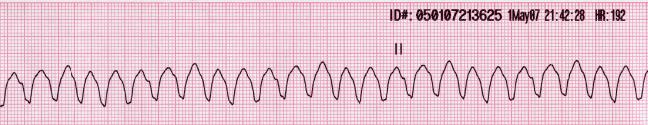
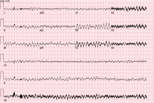
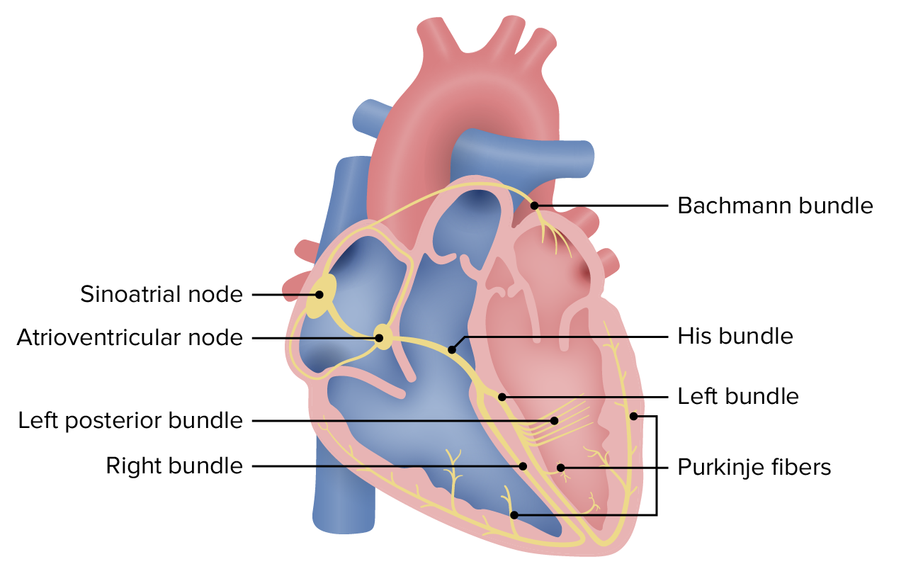
00:01 Welcome back to the ECG lecture series. 00:04 We are going to be speaking about ventricular arrhythmias in this series. 00:08 Remember that ventricular arrhythmias are those that occur below the AV node. 00:14 Remember when we talked about supraventricular arrhythmias, we mentioned arrhythmias that occurred in the atria above the AV node. 00:21 Now we're speaking about arrhythmia's abnormal electrical activity that occurs below the AV node and it is almost all of these ventricular arrhythmias that occur below the AV node have abnormal conduction through the ventricle so there's a broad QRS. 00:38 Usually more than 3 little boxes that's 0.12 seconds. 00:43 Also, the ST segments and the T waves go on opposite directions from that taken by the QRS and the beat looks very bizarre and abnormal compared to the underlying normal rhythm. 00:56 The QRS and ST segments of ventricular premature beats are very different in form from the normal sinus or regular beats as I'm going to show you in a moment. 01:07 So here is a typical premature ventricular contraction fondly known as a PVC, also called a VPB, a ventricular premature beat. 01:19 So, you'll hear both abbreviation: PVC or VPB. 01:23 Notice first of all, there's a wide QRS and it's directed upward in the blue box. 01:30 Notice that the ST segment in the green box is directed downward and in the opposite direction from the direction that the QRS went. 01:40 And also notice how different this beat is from the previous sinus beats. 01:46 There's two sinus beats there. Narrow QRS preceded by a P wave. 01:50 There's no P wave in front of this beat. 01:52 It's a wide bizarre-looking complex with a very bizarre-looking ST and T wave. 01:58 And that's because the depolarization and the repolarization are completely abnormal compared to the normal underlying rhythm. 02:05 Here we see three PVCs in a row called a triplet of PVCs. 02:10 You'll notice, first beat is a normal beat, P, QRS, and T. 02:16 The next beat, normal beat, P, QRS, and T. And then wow. 02:20 Look at that three big PVCs, tall P waves, abnormal ST and T waves segments, and then following that we're back to the normal rhythm: P, QRS, and you don't see the T there. 02:33 Here we see two different PVCs in the same patient. 02:38 So, notice in the green boxes, let's look -- the first beat is normal, P, QRS, and T. 02:44 And then there's a beat in purple in the green box. It a wide bizarre QRS. 02:51 They're differently oriented, completely oppositely oriented ST and T wave. 02:55 Then we return normal beat, P, QRS, T. Normal beat, P, QRS, T. Normal beat, P, QRS, T. 03:03 Next green box, another bizarre widely oriented QRS with the ST and the T wave going in the opposite direction from the direction of the PVC QRS. 03:18 So, notice in this patient, there is more than one focus of PVCs. 03:24 And so, this is called multifocal or multiple originating origins of impulse coming from different areas in the ventricle. 03:35 By the way, multifocal PVCs are more worrisome than when all the PVCs look the same.
About the Lecture
The lecture ECG of Premature Ventricular Contraction (PVC) by Joseph Alpert, MD is from the course Electrocardiogram (ECG) Interpretation.
Included Quiz Questions
Which of the following could be the location of depolarizing cells that are causing ventricular arrhythmias?
- Purkinje bundles
- Sinoatrial node
- Left atrium
- Right atrium
- Fossa ovalis
Which of the following is characteristic of PVCs?
- T-wave in the opposite direction of QRS
- Narrow QRS
- T-wave directed upward
- QRS directed downward
- Preceded by P waves
Author of lecture ECG of Premature Ventricular Contraction (PVC)

Joseph Alpert, MD
Customer reviews
5,0 of 5 stars
| 5 Stars |
|
5 |
| 4 Stars |
|
0 |
| 3 Stars |
|
0 |
| 2 Stars |
|
0 |
| 1 Star |
|
0 |
