Playlist
Show Playlist
Hide Playlist
ECG of Atrial Premature Beats

-
Slides Supraventricular Arrhythmias.pdf
-
Download Lecture Overview
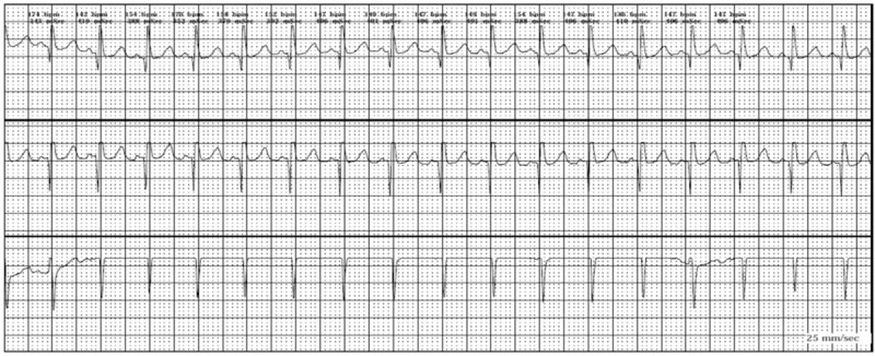
00:01 So let's start with the simplest atrial arrhythmias. These are single atrial premature beats. 00:07 You can have 1-3 of these beats sometimes in a row. They originate in the atrium. 00:13 They are extra beats therefore they're called premature atrial beats and they are often preceded by an abnormally appearing P wave but sometimes you can't see the abnormally appearing P wave. 00:26 It doesn't appear in the lead or the rhythm strip you're looking at. 00:30 So here is an example of atrial premature beats. Let's look at the first complex. 00:37 We don't see the P wave there. So, let's look at the second QRS. 00:42 There's a normal T wave then you have a normal P wave in the third beat and the T wave And, oh my goodness, look at that. There's a beat that occurs much too early. 00:53 Now if you look at the T wave there, you'll note and compare it to the previous T wave, you'll notice it's different. 01:00 Why? Because there's an abnormal P wave added into that T wave. 01:05 So, compare it to the T wave of the second beat. The T wave from the second beat is a normal T wave and the T wave of the third beat is a combination of the T wave plus the abnormal P wave from the premature beat. 01:18 Now if we look at the fifth beat, that's normal. 01:21 Normal P, normal QRS, normal -- not normal T. An abnormal T cuz again, it's the combination of the T wave and the premature beat and you see the premature beat. 01:31 Each of these two premature beats are marked with a green arrow and you can see in subsequently, this rhythm continues. Normal beat, premature beat. 01:40 Normal beat, premature beat. Normal beat, premature beat. 01:42 And in each case, the T wave before the premature beat is abnormal cuz it's the combination of a normal T wave and an abnormal P wave. 01:51 And that's pointed out here. The second hump on the T wave corresponds to the P wave from the atrial premature beat and it's partially hidden or combined within the T wave. 02:02 The QRS of the atrial premature beat is slightly widened because there's a little bit of intraventricular delay caused by the abnormal arrival of the premature beat's electrical impulse. 02:15 In other words, the system hasn't totally reset itself so you can have a slightly abnormal QRS but it's still narrow but not quite as narrow as the normal beat. 02:25 And that's probably because the whole system below the AV node hasn't had the chance to totally reset itself so you get a little tiny amount of block there but still narrow QRS.
About the Lecture
The lecture ECG of Atrial Premature Beats by Joseph Alpert, MD is from the course Electrocardiogram (ECG) Interpretation.
Included Quiz Questions
Which of the following is associated with an atrial premature beat pattern?
- Abnormally shaped P waves
- Often accompanied by symptoms such as palpitations or chest pain
- The beats originate in the AV node
- A wide QRS complex
- Prolonged P-R interval
Author of lecture ECG of Atrial Premature Beats

Joseph Alpert, MD
Customer reviews
5,0 of 5 stars
| 5 Stars |
|
5 |
| 4 Stars |
|
0 |
| 3 Stars |
|
0 |
| 2 Stars |
|
0 |
| 1 Star |
|
0 |
