Playlist
Show Playlist
Hide Playlist
ECG of Atrial Flutter and Fibrillation

-
Slides Supraventricular Arrhythmias.pdf
-
Download Lecture Overview
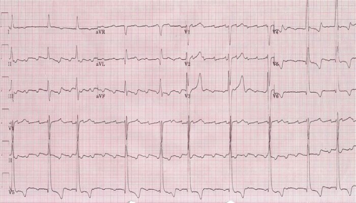
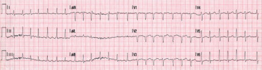
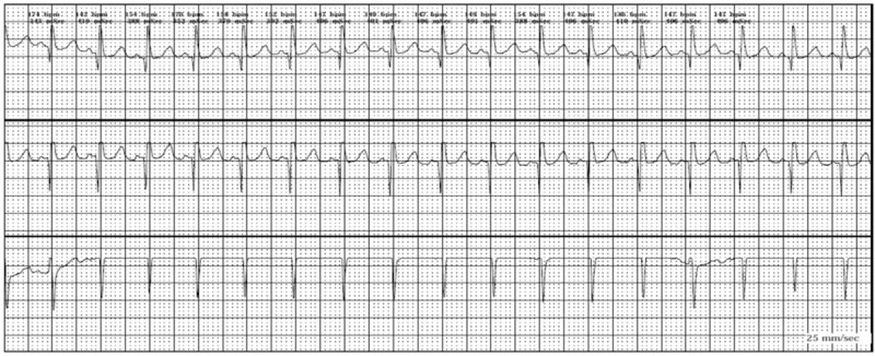
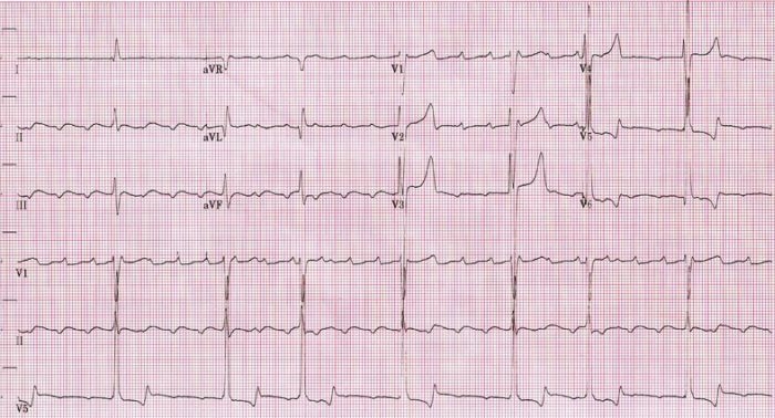
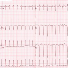
00:01 Another form of a supraventricular rhythm which is similar to the one we just saw but slightly different is called atrial flutter. 00:10 And again, this is a circus movement of depolarization in the atrium with every - often every third or fourth beat getting through the AV node causing a ventricular depolarization. 00:20 Let's see an example. Again, it's a circus movement, it usually goes at 300 per minute. 00:26 Often, 2:1 block so you get a ventricular rate of 150. And on the EKGs, the flutter waves have a sawtoothing appearance. 00:36 Looks like sawtoothing. 00:38 Here we see an example. Notice, this is a heart rate of 75, so that's - if you divide 300 down to 75 that's 4:1. 00:48 So, this is 4:1 flutter and you'll notice the beats occurring. 00:53 They're actually - there are some 4:1, and then sometimes it'll go to 3:1 and 2:1, it can go back and forth. 01:01 In this case it's 4:1. And look at the flutter waves there. 01:04 You see the sawtoothing in between the QRSs? If you see the first beat that's the QRS. 01:10 And then sawtooth, sawtooth, sawtooth, QRS, sawtooth, sawtooth, sawtooth, QRS, sawtooth and so forth. 01:19 You'll notice also this patient has a left bundle branch block. 01:25 They have a wide QRS, greater than three little boxes that preexisted the atrial flutter. 01:31 And again, we're in this pattern, the sawtoothing pattern means atrial flutter usually at 300 per minute. 01:39 You'll notice there's one big box between the saw teeth. 01:42 Here's another example. Here - beautiful flutter waves. 01:47 Notice, again, this is also a rate of - ventricular rate of 75, so it's 4:1 atrial flutter. 01:54 See how clear it is. Sometimes it's not quite so clear but in any case, this is another example of 4:1 AV block with atrial flutter. 02:04 And of course, the ventricular rate is 75 per minute. 02:08 Now, atrial fibrillation is not as organized. It's not a circus movement. 02:13 In fact, it's chaotic atrial activity in the atria. 02:16 I like to tell the patients with my hand. 02:19 So, this is a supraventricular tachycardia, it's going very fast and then periodically the ventricle goes. 02:25 But what happens in atrial fibrillation is this. 02:29 It's totally random and irregular; the number of beats that are getting through. 02:35 So, you have a completely irregular heart rhythm as opposed to atrial flutter which is regular or supraventricular tachycardia which is regular. 02:43 So atrial fibrillation again, the result of multiple chaotic depolarization waves in the atria. 02:50 Some of the waves get through the AV node in a random fashion leading to a tachycardia with a very irregular heart rate. 02:58 The QRS is usually narrow, again, unless we get to the same thing I mentioned before. 03:03 The heart rate is so fast that you get a so-called rate related right bundle. 03:08 And that occurs only when the QRSs are very close together. 03:12 So, here is atrial fibrillation with a reasonable heart rate, not too fast. 03:18 Notice, you - the distance between the two R - the R's of the QRS is completely random. 03:26 Some are short, some are long, some are medium. 03:30 So, this is called an irregularly irregular rhythm. It's completely irregular random. 03:37 And you can do that by - if you have a compass and you put it on the first QRS to QRS rate the next one, it's much shorter. 03:47 Oh, the next one is much longer. Oh, the next one is intermediate. Oh, it's much shorter. 03:51 So again, the next one much longer. 03:53 And you'll see little waves occurring there but they're not organized. 03:57 They're - they are fibrillation waves, not flutter waves. 04:02 So, the heart rate is more irregular than in flutter. 04:04 The waves seen between the QRS's are variable in size and shape. 04:08 They're not nicely sawtooth. There's no sawtoothing. 04:12 Here we see, again, this example of a normal ECG, normal QRSs, no hypertrophy, no myocardial infarct and a markedly irregular heartbeat. 04:23 Look at the bottom strip. You'll see how totally irregular that is. 04:27 You don't see sawtooth, you don't see abnormal P waves. This is atrial fibrillation. 04:32 You can tell it by the grossly irregular heart rate. 04:36 You can measure out the distances between the two R waves all the way through, every one of them is slightly different.
About the Lecture
The lecture ECG of Atrial Flutter and Fibrillation by Joseph Alpert, MD is from the course Electrocardiogram (ECG) Interpretation. It contains the following chapters:
- Atrial Flutter
- Atrial Fibrillation
Included Quiz Questions
Which of the following ECG findings is characteristic of atrial flutter?
- Sawtooth atrial waves
- Wide QRS complexes
- Irregularly irregular heart rhythm
- Irregular heart rate
- T-wave inversion
Which of the following ECG findings is characteristic of atrial fibrillation?
- Irregularly irregular heart rhythm
- Sawtooth atrial waves
- 3–4 P waves to 1 QRS complex
- Regular heart rate
- T-wave inversion
Which of the following is TRUE about atrial fibrillation?
- It is the result of multiple chaotic depolarization waves in the atrium.
- It is a circus wave of depolarization in the atrium.
- It is a circus wave of depolarization in the ventricle.
- It is the result of multiple chaotic depolarization waves in the ventricle.
- It is preceded by atrial flutter.
Author of lecture ECG of Atrial Flutter and Fibrillation

Joseph Alpert, MD
Customer reviews
5,0 of 5 stars
| 5 Stars |
|
5 |
| 4 Stars |
|
0 |
| 3 Stars |
|
0 |
| 2 Stars |
|
0 |
| 1 Star |
|
0 |
