Playlist
Show Playlist
Hide Playlist
ECG of Anterior Myocardial Infarction (MI)

-
Slides ECG of Myocardial Infarction.pdf
-
Download Lecture Overview
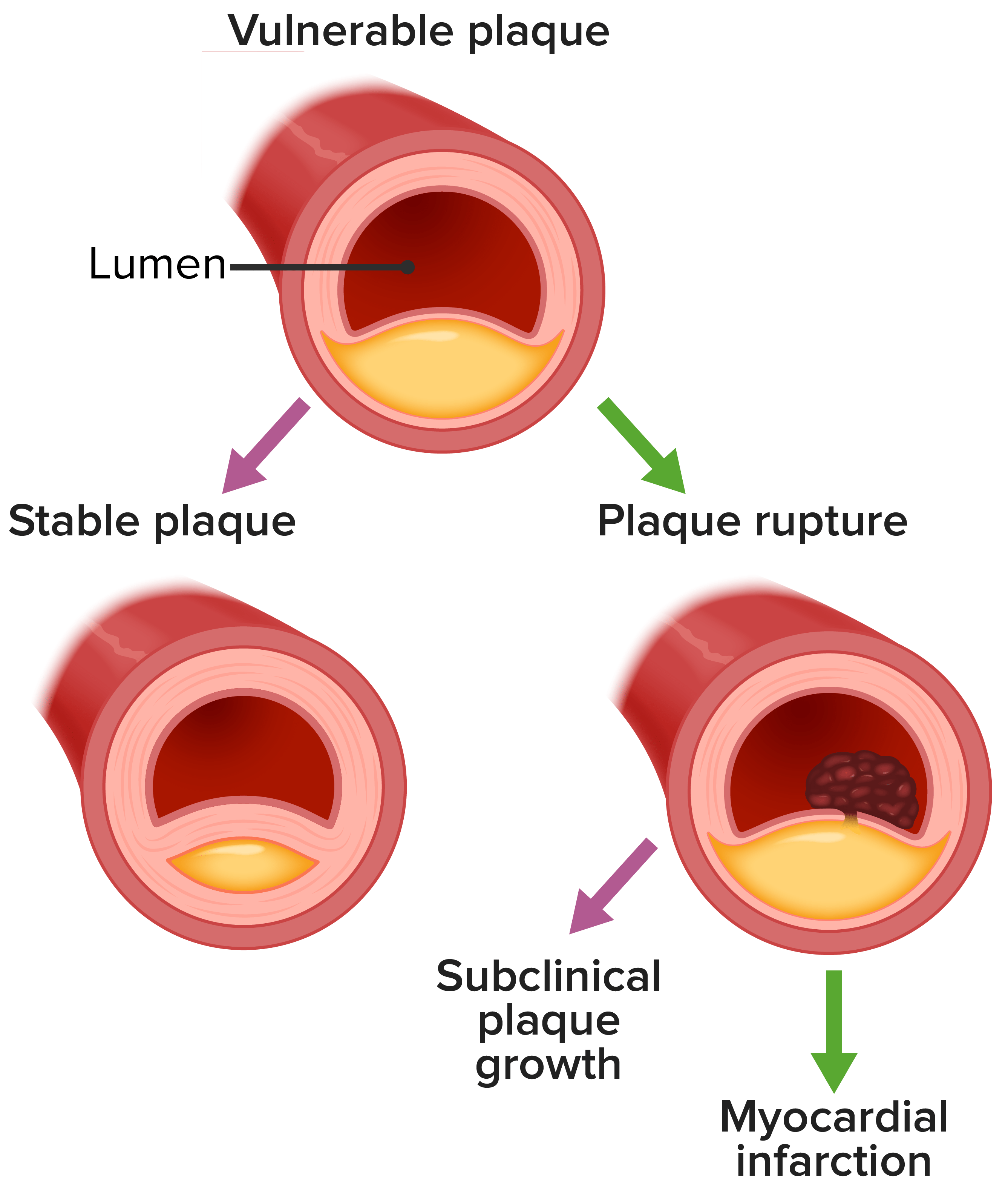
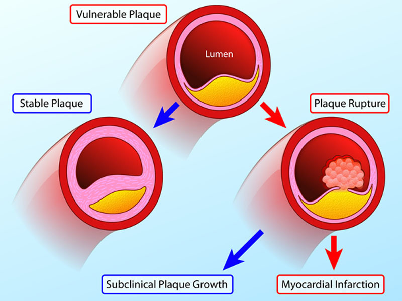
00:01 Here, we see an example of an anterior wall myocardial infarct. 00:05 Notice that there's ST elevation in all of the 6 precordial leads. 00:09 A little bit in V1 and a little in V6 but really very impressive huge ST elevation in leads V2, V3, V4, and even pretty impressive in V5. 00:23 These very high ST elevations almost 10 mm in size. 00:29 These are sometimes been called tombstone changes. Why? This is a very, very dangerous myocardial infarct. It's an occlusion. 00:38 Very likely proximal of the left anterior descending coronary artery. 00:42 A huge amount of heart muscle is threatened. This is a real big emergency. 00:47 Off to the cath lab as fast as possible and open up the LAD again. 00:51 So, this is a very nasty anterior MI. 00:55 Here's another one not quite so nasty. 00:59 You'll notice ST elevation in leads V1, V2, and V3 and also in aVL so that suggests that there's a certain lateral component to this. 01:11 So possibly, this is a LAD that has branches in the lateral wall or it may be even an involvement with the left circumflex and the left anterior descending. 01:24 It may be that for example a patient had a previous narrowing of the circumflex and when the left anterior descending goes down, there's lack of good blood flow down the circumflex as well. 01:36 So, in any case, this is another example of anterior MI. 01:41 Also, what's of interest, notice the QRS. Look at the QRS particularly in leads V1, V2, and partly in V3. 01:51 There's a Q wave. The QRS starts with a Q wave. 01:54 Starts with a hole in the heart that's electrically gone and you'll notice also that there's a QS pattern in lead aVL. 02:05 Same thing that this is telling you that there's myocardium that's no longer depolarizing. 02:11 Again, we point that out. 02:14 There are Q waves in leads V2, V3, and aVL and I think if we magnify that enough, there's a little tiny Q wave also in lead V3. 02:23 So here's some unknowns. Look at this ECG. I'll tell you a clue to start with. 02:30 There's a myocardial infarct here. Tell me where it is. 02:41 Is this an acute inferior wall MI? The answer is yes. ST elevation in leads 2, 3, and aVF. 02:48 Also, of interest, notice right at the very beginning there's a standardization. 02:54 The box that's ten points high telling you that this is the right standardization for this ECG. 03:01 Let's look at another one. Take a look at the next one. I'll also tell you this is an MI. 03:09 You tell me where it is. 03:17 Is this an acute anterior? Yes. There's ST elevation in leads V2, V3, V4, and V5. 03:26 So again, acute anterior, that's LAD--left anterior descending artery. 03:31 Acute inferior, usually the right coronary but can be in 10% of people of left circumflex. 03:38 Here, we see the ST elevation arterially.
About the Lecture
The lecture ECG of Anterior Myocardial Infarction (MI) by Joseph Alpert, MD is from the course Electrocardiogram (ECG) Interpretation.
Included Quiz Questions
ST elevations in leads II, III, and aVF would be most indicative of which type of MI?
- Acute inferior wall MI
- Acute superior wall MI
- Acute lateral wall MI
- Acute anterior wall MI
- Acute septal MI
ST elevations in leads V2, V3, V4, and V5 would be most indicative of which type of MI?
- Acute anterior wall MI
- Acute inferior wall MI
- Acute superior wall MI
- Acute lateral wall MI
- Acute septal MI
Author of lecture ECG of Anterior Myocardial Infarction (MI)

Joseph Alpert, MD
Customer reviews
5,0 of 5 stars
| 5 Stars |
|
2 |
| 4 Stars |
|
0 |
| 3 Stars |
|
0 |
| 2 Stars |
|
0 |
| 1 Star |
|
0 |
An excellent approach to ECGs. Makes a difficult topic very simple to understand.
very simple and clear explanation and repetition and testing help a lot. thannk you
