Playlist
Show Playlist
Hide Playlist
Carpal Tunnel

-
Slide Carpal Tunnel.pdf
-
Download Lecture Overview
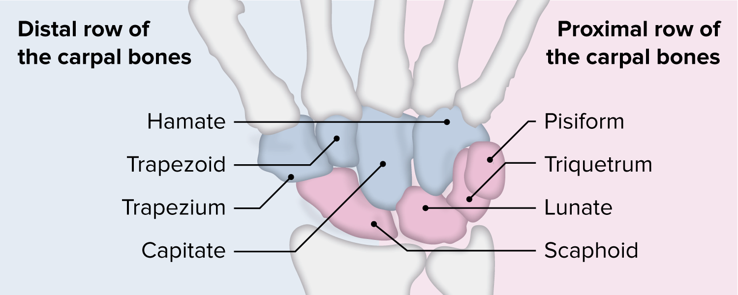
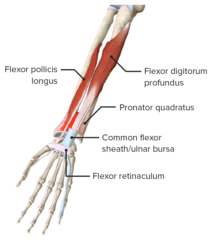
00:01 So, I've mentioned it a few times before in the previous section. 00:04 But now, we're going to look at the carpal tunnel, an important passageway that allows structures to pass from the forearm into the hand. 00:13 So, the carpal tunnel is situated really over the wrist. We can see that it's formed by the flexor retinaculum. 00:20 So, this is the roof. We're looking at the carpal tunnel in cross-section. 00:24 So, we've cut a section as the diagram in the top of the screen indicates and we're now looking through it as if we're looking into the digits of the hand. 00:33 So, the roof of it is formed by the flexor retinaculum. 00:37 The base or the floor of the carpal tunnel is formed by these series of bones forming the carpal groove. 00:44 Here, we can see we've got the pisiform. 00:46 So, we can see this bone is forming the boundary of the carpal tunnel alongside the hook of the hamate. 00:52 And these two bones form the medial boundary. We can also see on the lateral aspect, we've got the tubercle of the scaphoid bone, tubercle of trapezium. 01:02 And these form the lateral boundary. 01:05 So, what we have is a medial and lateral boundary of the carpal tunnel, a roof, and a floor. 01:13 If we then look at what we find inside the carpal tunnel, then, we can see a whole series of those structures that we spoke about in the forearm that are now passing towards the hand. 01:22 We spoke about their attachments on the digits and this carpal tunnel allows those tendons to pass from the muscle bellies located in the forearm and pass their way through the carpal tunnel, onto their insertion points on the digits. 01:38 So, here, we could see the flexor digitorum profundus tendons. 01:40 So, they're deep within the carpal tunnel. Sitting on top of them or sitting more superficial, we have the tendons of flexor digitorum superficialis. 01:49 So, here, we can see a whole series of tendons, eight in total, passing from the forearm towards the hand through the carpal tunnel. We also have the tendon of flexor pollicis longus. 02:00 We have the median nerve and then, associated of all of these tendons, we have those synovial sheats. 02:07 Exactly the same as what we had on the extensor aspect and these help to prevent friction. 02:12 So, a mixture of tendons and nerves we can see here. 02:16 What doesn't pass through the carpal tunnel is the ulnar nerve and the ulnar artery. 02:21 We can see these running separately to the carpal tunnel and these are running in a channel called Guyon's canal. 02:29 So, they don't run within the carpal tunnel. That's important to recognize. 02:33 Palmaris longus tendon also passes more superficially within this space and that's passing most superficial underneath the flexor retinaculum to go and form the apex of the palmar aponeurosis.
About the Lecture
The lecture Carpal Tunnel by James Pickering, PhD is from the course Anatomy of the Hand.
Included Quiz Questions
Which structures pass through the carpal tunnel? Select all that apply.
- Tendons of flexor digitorum profundus
- Ulnar artery
- Tendons of flexor digitorum superficialis
- Tendon of flexor pollicis longus
- Median nerve
Author of lecture Carpal Tunnel

James Pickering, PhD
Customer reviews
5,0 of 5 stars
| 5 Stars |
|
5 |
| 4 Stars |
|
0 |
| 3 Stars |
|
0 |
| 2 Stars |
|
0 |
| 1 Star |
|
0 |
