Playlist
Show Playlist
Hide Playlist
Cardiac Case: 22-year-old Woman with Leg Edema, Dyspnea and Fatigue

-
Cardiac Case 22-year-old Woman with Leg Edema Dyspnea and Fatigue.pdf
-
Download Lecture Overview
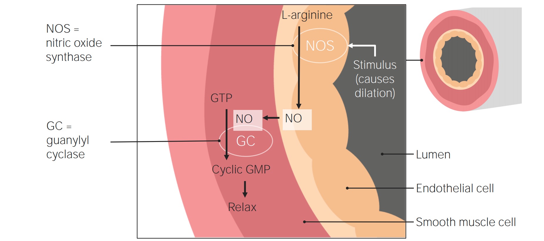
00:01 A 22-year-old graduate student is seen in clinic because of increasing leg edema that has become refractory to diuretics. 00:07 She was given a diuretics by her primary care physician and it was thought just to be some, perhaps some edema from her menstrual cycle. 00:17 She also has noted though, much more worrisome, increasing dyspnea on exertion and fatigue and previously, she was healthy. 00:25 It's interesting that her mother has been diagnosed with systemic lupus erythematosus so often, this is found in families where there's a lot of autoimmune diseases. 00:36 Physical exam shows blood pressure's pretty normal for young female, 104/78, heart beat a little bit elevated at 86, and the peripheral oxygen saturation is abnormal, 88% on room air suggesting lung disease. 00:50 And her jugular venous pulse is also elevated, should be under 10 and particularly way under 10 for such a young woman. 00:58 The lungs are clear and the cardiac exam shows a loud pulmonic component to the second heart sound but no murmurs. 01:06 So, let's think about that. Here's normal, lub-dub, lub-dudub, lub-dudub, lub-dub. 01:12 And the secondary component, second splitting of the second heart sound is the pulmonic component, lub-dudub, lub-dudub, lub-dudub. There you can see it's accentuated. 01:22 You can hear it at the apex where you normally don't hear the splitting. 01:26 That tells you that P2, the pulmonic component is increased. 01:30 That suggest pulmonary hypertension and she has 2-3+ peripheral edema. 01:34 So the critical factors here in the history of course, dyspnea, unusual in a young woman like this. Hypoxemia suggesting lung disease. 01:42 Elevated jugular venous pressure suggesting a right heart failure. 01:47 The loud P2 suggesting pulmonary hypertension and the peripheral edema suggesting some right ventricular failure. 01:55 And here's her ECG. It shows right ventricular hypertrophy. 02:00 Again, notice the large R wave in V1. You're supposed to have only a small R wave in V1. 02:05 And there's right axis deviation so again, we are dealing with somebody with cor pulmonale. 02:13 That is with a right ventricle that's been over strained, likely pulmonary hypertension. 02:19 The chest X-ray shows right ventricular dilatation and an echocardiogram shows reduced right ventricular systolic function and the pulmonary artery systolic pressure is estimated at a frightening 96 mmHg. 02:33 In other words, the same level as her arterial blood pressure so she has arterial blood pressure in her pulmonary circuit. 02:42 Very severe pulmonary hypertension, she has a CT pulmonary angiogram and there's no pulmonary emboli This could one of the causes of pulmonary hypertension. 02:51 So the diagnosis is idiopathic meaning unknown cause of pulmonary hypertension with RV failure. 02:58 It's thought to be, in some form, genetics and might be related to the fact that her mother had lupus erythematosus. 03:07 It's another form of cor pulmonale, the first form we saw earlier was due to cigarette smoking and damage to the lungs. 03:15 This one is due to damage to the very small blood vessels in the lung. 03:19 It's also called primary pulmonary hypertension or pre-capillary idiopathic pulmonary hypertension. 03:26 The little pulmonary capillaries are obliterated. 03:29 There's severe vasoconstriction in the lung and there's also dysfunction of the pulmonary endothelium that is the lining of the small blood vessels. 03:38 And pulmonary angiography is these patients demonstrates a lung vasculature that looks like a pruned tree where the leaves have been cut off. 03:46 This used to be untreatable but these days we have some vasodilating drugs that do help. 03:53 Endothelin blockers, prostacyclin, phosphodiesterase-type inhibitors, that's like sildenafil, Viagra that also dilates the blood vessels in the lung, and there's a whole bunch of others that are currently being investigated. 04:07 The major treatment if she doesn't respond to medicines would be lung or heart/lung transplantation. 04:13 And then of course in the future, we're expecting new therapies for this condition all the time. 04:20 The prognosis for this condition was formerly very poor. 04:24 It's much improved with the new drugs and with the possibility for heart or heart/lung transplantation.
About the Lecture
The lecture Cardiac Case: 22-year-old Woman with Leg Edema, Dyspnea and Fatigue by Joseph Alpert, MD is from the course Cardiovascular Cases.
Included Quiz Questions
A 22-year-old graduate student presents to the clinic because of increasing leg edema that has become refractory to the diuretics prescribed by her primary care physician. She also notes increasing dyspnea on exertion and fatigue. She has been previously healthy. Her family history indicates that her mother has a diagnosis of systemic lupus erythematosus. On physical examination, her blood pressure is 104/78 mm Hg, heart rate is 86 beats/min, peripheral oxygen saturation 88% on room air, and the jugular venous pulse is 12 cm of water. Lung auscultation shows no abnormalities. She has 2-3 pitting edema on her lower extremities. Which of the following would you find in this patient?
- Loud pulmonic component to the second heart sound
- Loud aortic component to the second heart sound
- Opening snap at the apex
- Paradoxical splitting of the second heart sound
- Systolic ejection murmur, radiating to the carotids
A 22-year-old graduate student presents to the clinic because of increasing leg edema that has become refractory to the diuretics prescribed by her primary care physician. She also notes increasing dyspnea on exertion and fatigue. She has been previously healthy. Her family history indicates that her mother has a diagnosis of systemic lupus erythematosus. On physical examination, her blood pressure is 104/78 mm Hg, heart rate is 86 beats/min, peripheral oxygen saturation 88% on room air, and the jugular venous pulse is 12 cm of water. On lung auscultation, her lungs are clear. On extremities examination, 2–3 peripheral edema is noted. After several diagnostic studies, a diagnosis of idiopathic pulmonary hypertension is reached. Which of the following is the best initial treatment for this patient?
- Phosphodiesterase 5 inhibitors
- Angiotensin-converting enzyme inhibitors
- Loop diuretics
- Lung transplantation
- Lung and heart transplantation
Author of lecture Cardiac Case: 22-year-old Woman with Leg Edema, Dyspnea and Fatigue

Joseph Alpert, MD
Customer reviews
5,0 of 5 stars
| 5 Stars |
|
1 |
| 4 Stars |
|
0 |
| 3 Stars |
|
0 |
| 2 Stars |
|
0 |
| 1 Star |
|
0 |
CARDIOLOGY MADE EASY HE MADE IT EASY HE IS THE MAN WITH A PLAN RIGHT HERE
