Playlist
Show Playlist
Hide Playlist
Brachial Plexus

-
Slide Brachial Plexus.pdf
-
Download Lecture Overview
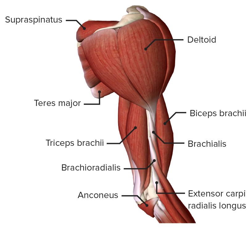
00:01 So, now, let's look at the brachial plexus. 00:04 The brachial plexus is a complex collection of nerves within the axilla. 00:10 It serves to allow new logical structures to connect the neck to the upper limb. 00:15 And that's important to allow the wide range of function of the various muscles within the upper limb. 00:22 It's important also for sensation cutaneously of the skin. 00:29 So, the brachial plexus, like I said, is a connection of neurological structures that leave the spinal cord and pass laterally from the neck region into the axilla. 00:38 The main substance of the brachial plexus as we can see here is wedged in between two important muscle bellies. 00:44 We'll come to those in a moment. What you can see though are the roots of the brachial plexus. 00:49 So, these are running away from the spinal cord, containing both motor and sensory fibers. 00:55 There were also connections to the main sympathetic trunk that's running down lateral to the vertebrae and this is the sympathetic trunk's gray rami communicantes. 01:08 And these are important as they allow sympathetic fibers to piggyback onto these roots of the brachial plexus, taking sympathetic innervation down to the upper limb. 01:20 That's important for piloerection of the hairs on your arms for sweat glands, etc. 01:24 Importantly though, there are no parasympathetic fibers running cutaneously towards the upper limb. 01:31 And this is similar for the lower limb as well. 01:35 As I mentioned, the brachial plexus is wedged in between two important muscle bellies. 01:38 Posteriorly, we have the middle scalene muscle and anteriorly, we have the anterior scalene muscles which you can see here. 01:47 In close association in coming up to meet the brachial plexus from inferiorly, we have the subclavian artery. 01:53 And here, in this view, you can see the subclavian artery running laterally out of the chest wall, combining, merging with the brachial plexus between those two scalene muscle bellies as they pass into the axilla via the cervicoaxillary canal. 02:09 Once it's in that canal, it then enters the axilla by passing deep to the clavicle which we can see here. 02:15 And here's a reminder, the location of the first rib and scapula. 02:21 The brachial plexus has an important number of parts. 02:23 And they start from the spinal cord located at C5 all the way through to T1. 02:30 So, we can see it coming out of the spinal cord, the roots of the brachial plexus. 02:36 Here, we have C5, C6, C7, C8, and then, finally, T1. So, we have five roots of the brachial plexus. 02:46 And as you can see here in this diagram, these roots to some extent, combine and in some other instances, they don't. 02:54 So, the formation of trunks from these roots takes a very specific pattern. 03:00 So, here, we can see the formation of the superior trunk by the C5 and C6 roots uniting. 03:09 So, this occurs as you move laterally away from the vertebral column which is containing the spinal cord. 03:16 We see the formation of C5, C6 roots converging to form the superior trunk. 03:22 The seventh cervical spinal root comes out and it doesn't combined of anything. 03:27 It stays on its own and it forms what's known as the middle trunk. 03:31 Then, most inferiorly, we have the inferior trunk formed by C8 and T1. 03:37 So, we can see from the five roots, we now have three trunks. 03:42 Coming away from each of these trunks is a number of divisions. And we have six divisions in total. 03:50 That means each of these three trunks give rise to two divisions. 03:56 And these are known as anterior and posterior divisions. 04:00 So, here, we can see the superior trunk giving rise to its anterior and posterior division. 04:06 The same applies for the middle, giving rise to its anterior and posterior division. 04:12 And then, finally, inferior trunk gives rise to its anterior and posterior division. 04:20 So, we've gone from five roots, C5, C6, C7, C8, T1 to three trunks. 04:27 By C5 and C6 forming the superior trunk, C7 continuing as the middle trunk, and then, C8 and T1 combining to form the inferior trunk. 04:39 Each of these trunks then gives rise to either an anterior or a posterior division. 04:45 And here, we can see each of those three trunks giving rise to their two divisions. 04:51 The main part of the brachial plexus that you really should be familiar with are the kind of terminal cords that are coming away from the brachial plexus. 04:59 And this is really formed by these anterior divisions uniting or in some instances, not uniting and running on their own. 05:07 And the ultimate combination of the posterior divisions coming together. 05:12 So, here, we can see, we have the lateral cord. 05:16 The lateral cord is formed by the two anterior divisions that are coming from the superior and middle trunks, giving rise to the lateral cord. 05:26 So, where we can see the superior and middle trunks anterior divisions, they're combining to form the lateral cord. 05:34 Where we find all three posterior divisions, so, the one from the middle, one from inferior, and one for middle, all of those converge together to form the posterior cord. 05:48 That only leaves the anterior division of the inferior trunk and that gives rise to the medial cord. 05:57 So, we've gone from five roots, C5, C6, C7, C8, T1. C5 and C6 have combined to form the superior trunk. 06:11 C8 and T1 combine to form the inferior trunk. C7 has remained on its own as the middle trunk. 06:18 So, five roots down to three trunks. Each of those trunks then gives rise to an anterior and posterior division. 06:27 With the anterior divisions of superior and middle forming the lateral trunk, the anterior division of the inferior trunk forming the medial cord, and the three posterior divisions of each of the superior, middle, and inferior trunks forming the posterior cord. 06:47 This is quite a complicated network of interconnecting nerves and it's really helpful if you just have blank sheets of paper and draw this out for yourselves. 06:56 It's not that complicated once you start getting into repetition in doing it. 07:01 But it's an important structure you need to familiarize yourself with. 07:05 As a reminder, here, we can see the formation of the lateral cord. 07:08 Here, we can see the formation of the posterior cord. 07:12 And finally, we can see the formation of the medial cord. 07:18 So, the formation of the lateral cord comes from the two anterior divisions from the superior and middle trunks. 07:26 So, passing laterally, the superior and the middle trunks, each give rise to an anterior division and those anterior divisions form the lateral cord. 07:36 So, now, let's carry on looking at the brachial plexus and a number of important terminal nerves that come away from it. 07:43 And from these, there are a number of branches, and you'll need to check with your own curricular whether you need to know all of these branches because there are quite a lot of small branches. 07:53 So, let's have a look and remind ourselves of the lateral, medial, and posterior cord. 07:58 The posterior cord here is highlighted slightly because it sits behind the axillary artery. 08:04 So, here, we can see the formation of a number of terminal nerves. 08:09 So, let's have a look at the lateral cord. And the main nerve coming away from the lateral cord is the muscular cutaneous nerve. 08:16 We'll come across that in a moment or two when we look at the nerves of the arm. 08:21 But the lateral cord principally gives off the terminal nerve which is the musculocutaneous nerve. 08:27 It also gives rise to what's known as the lateral root of the median nerve. 08:32 And the median nerve is the second terminal nerve coming away from the brachial plexus and it's formed from a contribution of both the lateral and the medial cord. 08:42 From the lateral cord, we have the lateral root of the median nerve. 08:47 And from the medial cord, we also have this medial root of the median nerve. 08:52 So, the median nerve is formed from both the lateral cord and the medial cord. 08:58 The medial cord then carries on, on its own as the ulna nerve, forming the third of these terminal nerves. 09:07 The muscular cutaneous coming from the lateral, the ulna coming from the medial, and then, the median nerve coming from contributions of both the lateral and the medial cords by way of lateral and medial roots respectively. 09:24 So, it's a very complex picture coming away from the cords of the brachial plexus to form these terminal nerves. 09:32 What about the posterior cord? Well, the posterior cord which we can see here now running posterior to the axillary artery, that gives rise to the radial nerve as it continues down through the brachial plexus and then, passes to the posterior aspect of the arm. 09:47 And it also gives rise to an important nerve which is the axillary nerve. 09:51 And that passes away posteriorly towards the deltoid region. 09:55 So, a number of important terminal nerves that are coming away from the cords formed within the brachial plexus. 10:03 Let's have a look at this in slightly different perspective now. 10:07 So, here, we're looking at the posterior aspect of the brachial plexus. 10:12 So, we have the middle scalene muscles removed. 10:15 And you can see, again, the formation of the brachial plexus here coming away from the C5 brachial plexus root, we've got the dorsal scapular nerve and we can also see that passing now posteriorly onto the back musculature, the rhomboid muscles. 10:33 And here, we can see for detail the middle scalene muscle that it's passing through. 10:38 And here, we can see the rhomboid minor and rhomboid major muscles. 10:42 We can also see coming away from this C5 aspect of the brachial plexus, the most superior roots, we have its contribution to the phrenic nerve. 10:53 Remember, C3, 4, 5 keeps the diaphragm alive. That's the phrenic nerve. 10:58 And here, we see its contribution from that C5 root. 11:02 We can then continue and see the long thoracic nerve and the long thoracic nerve runs along the lateral aspect of serratus anterior which we can see here. 11:12 And you should be able to identify that on the lateral aspect of that muscle. 11:17 So, now, let's carry on and start looking at various other nerves that are coming away from the brachial plexus. 11:22 Here, we have the suprascapular nerve. 11:25 Remember, the suprascapular nerve goes to supply various muscles of the rotator cuff. 11:30 So, here, we have supraspinatus and infraspinatus muscles and the suprascapular nerve is passing through that subsuprascapular notch that we've spoken about in previous lectures. 11:43 If you remember on the anterior surface of the chest wall just underneath the clavicle, we have the subclavius muscle and here, we can see nerve to subclavius. 11:54 So, a whole series of these nerves that are passing very much from the roots and the trunks of the brachial plexus, a number of important nerves that are passing through to supply important structures within the upper limb and on the chest wall and in regard to the phrenic nerve within the diaphragm. 12:11 So, now, let's carry on and work our way through the brachial plexus giving rise to a number of nerves coming off it as it passes within the axilla. 12:20 So, here, we have the lateral cord which is giving rise to the median nerve. 12:25 You can see its lateral roots of the median nerve there coming from the lateral cord. 12:30 And again, here, you can see the muscular cutaneous nerve. 12:33 So, there's two main branches coming away from the lateral cord, the lateral roots forming the median nerve and the musculocutaneous nerve, supplying the muscles on the anterior aspect of the arm. 12:45 It also gives rise to some important structures within the substance really of the axilla, passing through the fatty mass of the axilla. 12:53 And here, we have the lateral pectoral nerve going to supply pectoralis minor and it's running alongside the thoreco-acromial artery which we saw previously. 13:03 Here, we see pectoralis major has then just been added to the anterior aspect of this image. 13:10 Here, we can see the medial cord. The medial cord is giving rise to the median nerve alongside the lateral cord via its medial root. 13:19 So, the formation of the median nerve, remember, contributed to by both the lateral and the medial cord. 13:25 Here, we can see its medial root forming that median nerve. 13:30 And the other main terminal nerve coming away from the medial cord we can see here is the ulnar nerve. 13:36 Coming away from the medial cord where we have the lateral pectoral nerve, coming away from the lateral cord, we now have the medial pectoral nerve coming away from the medial cord. 13:49 And you can see, there's various communicating branches with the lateral pectoral nerve which you can see here, forming another important complex of nerve, allowing essentially, a fair bit of redundancy. 13:59 So, some of these nerves become damaged. 14:01 They're able to be picked up by their connecting branches. Supplying pectoralis minor, we can see here. 14:08 Running down from the medial cord, we have the medial antebrachial cutaneous nerve. 14:13 So, antebrachial really means the forearm. So, this is supplying the medial aspect of the forearm's skin. 14:21 And here, we see the medial brachial cutaneous nerve supplying the medial aspect of the skin of the arm. 14:28 So, antebrachial, the forearm aspect, and the brachial being the arm aspect. 14:34 And here, we have these medial branches supplying them. 14:38 Now, let's have a look at the posterior cord. And here, we can see a view of the scapula. 14:44 So, we've had the anterior view of the scapula. 14:47 We've got the anterior surface with subscapularis muscle within it and we've had the chest wall removed. 14:54 So, we're looking into the axilla without the ribs there. 14:56 Running underneath the clavicle, we can see the posterior cord, associated on the top of the screen with the subclavian artery. 15:04 Here, we have the posterior cord. 15:06 The axillary artery's being removed to enhance clarity so you can see it better. 15:12 Coming off the posterior cord, we have the axillary nerve that's passing out to the posterior aspect of the axilla like we've mentioned through that quadrangular space. 15:22 You can see the two triangular spaces, triangular space, triangular interval on that diagram as well. 15:27 But moving through the quadrangular space, we have the axillary nerve. 15:31 And then, passing through the triangular interval, we have the radial nerve which you can see there. 15:38 If we add on some additional nerves to supply the subscapularis muscle, we have the superior and inferior subscapular nerves. 15:47 A whole series of nerves here associated with that musculature. 15:50 And then, finally, we have the thoracodorsal nerve which is innervating latissimus dorsi muscle. 15:57 Also surrounding this region, we have teres major, another muscle that's not associated with the rotator cuff but it's within this region and we can see, we have teres major muscle in this location. 16:10 These are all motor branches really that are supplying the substance of the musculature within this region. 16:15 And we also have a cutaneous branch. This one is the posterior brachial cutaneous nerve. 16:21 So, that is supplying the posterior aspect of the skin of the arm and we can see that passing away from this region as well. 16:29 So, there's a large number of nerves that are passing away from the brachial plexus and the brachial plexus in itself is an incredibly complicated structure. 16:38 So, make sure you refer to your own learning objectives and curricular to work out exactly what you need to know in both the formation of the brachial plexus, also, the branches that are coming away from the brachial plexus, and then, look at the gateways lecture to realize how lots of these terminal branches make their away from the axilla to their terminal structures.
About the Lecture
The lecture Brachial Plexus by James Pickering, PhD is from the course Anatomy of the Axilla.
Included Quiz Questions
Which statements concerning the axilla are correct? Select all that apply.
- The anterior wall is formed by the pectoralis major and minor.
- The medial wall is formed by the latissimus dorsi.
- The lateral wall is formed by the humerus.
- The posterior wall is formed by the subscapularis, teres major, and latissimus dorsi.
Which muscle forms part of the posterior wall of the axilla?
- Subscapularis
- Teres minor
- Infraspinatus
- Supraspinatus
- Serratus anterior
Which 5 ventral rami normally form the brachial plexus?
- C5, C6, C7, C8, and T1
- C2, C3, C4, C5, and C6
- C3, C4, C5, C6, and C7
- C4, C5, C6, C7, and C8
- C6, C7, C8, T1, and T2
The axillary nerve is a branch from which part of the brachial plexus?
- Posterior cord
- Medial cord
- Middle trunk
- Lateral cord
- Upper trunk
Which root forms the middle trunk of the brachial plexus?
- C7
- C5 and C6
- C6
- C8
- C8 and T1
Which cords of the brachial plexus form the median nerve?
- Lateral and medial
- Lateral
- Medial
- Posterior
- Posterior and medial
Which roots form the radial nerve?
- C5, C6, C7, C8, and T1
- C8 and T1
- C5 and C6
- C6 and C7
- C7 and C8
Author of lecture Brachial Plexus

James Pickering, PhD
Customer reviews
5,0 of 5 stars
| 5 Stars |
|
5 |
| 4 Stars |
|
0 |
| 3 Stars |
|
0 |
| 2 Stars |
|
0 |
| 1 Star |
|
0 |
