Playlist
Show Playlist
Hide Playlist
Boundaries of the Nasal Cavity
-
Slides Anatomy Nose.pdf
-
Download Lecture Overview
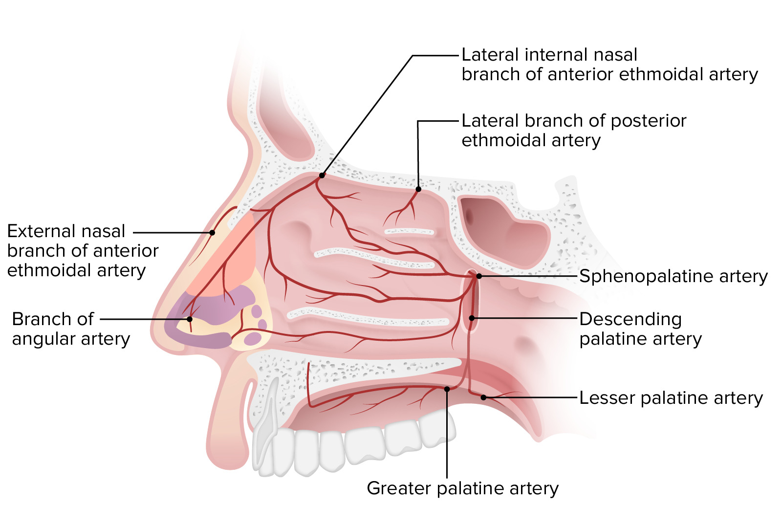
00:01 The area just inside the nostril that leads into the nasal cavity. 00:06 This area is called the nasal vestibule. 00:09 It's supported by the cartilage of the nose, and it's lined with tissue that contains small coarse hairs. 00:16 These hairs help to filter dust, sand and other particles that keep them from entering more deeply into the respiratory passageways depicting the nasal vestibule and the nasal cavity. 00:29 The nasal vestibule is shown right in through here, and then the nasal cavity is this larger area that we see posterior to the nasal vestibule as well as superior. 00:40 We have a hash green line right in through here. 00:43 And this is the Liman nasi. 00:45 The Limen nasi is the boundary line between the bony and the cartilaginous portion of the nasal cavity. 00:54 The nasal cavity proper and the vestibule meet right at the limen nasi so again the vestibule is here and then the nasal cavity lies above and then posterior. 01:05 Lastly, the limen nasi marks the transition of epithelial types. 01:11 Specifically we have a transition from squamous epithelium to respiratory epithelium. 01:18 The nasal septum again will divide the nasal cavity into a right nasal cavity and a left nasal cavity. 01:24 So this midline structure unless it's deviated, is shown in through here has a cartilaginous portion and it has a skeletal portion. 01:35 The skeletal portion of the nasal septum is in this superior view is the perpendicular plate of the ethmoid bone. 01:45 This bony contribution to the nasal septum is the vomer. 01:52 And then we have the septal cartilage, forming the anterior portion of the nasal septum. 01:59 Here we're looking at the lateral wall of the nasal cavity in the structures that form the right lateral wall that we're looking at. 02:07 will be duplicated on the left lateral wall as well. 02:11 First contribution to the lateral wall is the maxillary bone that we see in through here. 02:18 And then here we have the superior nasal concha associated with the ethmoid bone. 02:25 This projection is the middle nasal concha. 02:27 It's also an osteologic feature of the ethmoid bone and then existing as its own bone is the inferior concha, nasal concha. 02:38 And then lastly we have the palatine bone forming the lateral wall of this specific part of the palatine is the perpendicular plate. 02:47 But this extension vertically of the palatine is the perpendicular plate of the palatine bone forming the lateral wall. 02:59 Next to the nasal cavity is limited superiorly by a roof in the structures that contribute to the roof or the frontal dome. 03:10 This is more superior aspect of the frontal bone with the frontal sinus. 03:14 We also have a contribution from the nasal bone to the roof. 03:24 And the cribriform plate of the ethmoid contributes to the roof as well. 03:33 This is looking at the floor of the nasal cavity. 03:39 And so here we have the palatine process of the maxilla helping to form the floor. 03:45 This is also forming the hard palate anteriorly, horizontal plate of the palatine bone forming the posterior aspect of your hard palate as well as the floor. 04:02 And then we have on both sides here associated with the palatine processes are the maxillary bone. 04:09 We have a little opening here called the incisive canal and this is a passageway for the nasal palatine nerve and it also serves as a point of connection called an anastomosis between the greater palatine and the spenopalatine arteries.
About the Lecture
The lecture Boundaries of the Nasal Cavity by Craig Canby, PhD is from the course Head and Neck Anatomy with Dr. Canby.
Included Quiz Questions
Which of the following is a component of the roof of the nasal cavity?
- Frontal bone
- Palatine bone
- Superior nasal concha
- Maxillary bone
- Inferior nasal concha
Customer reviews
5,0 of 5 stars
| 5 Stars |
|
5 |
| 4 Stars |
|
0 |
| 3 Stars |
|
0 |
| 2 Stars |
|
0 |
| 1 Star |
|
0 |


