Playlist
Show Playlist
Hide Playlist
Bony Pelvis

-
Slides Bony Pelvis.pdf
-
Download Lecture Overview
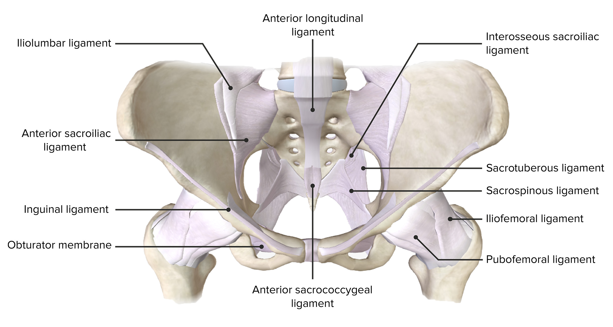
00:01 In this topic, we're going to have a look at the bony pelvis and the muscles of the pelvic floor. 00:08 So let's start off by reminding ourselves of the bones that form the bony pelvis or the pelvic girdle, which we can see here. We've got three bones. 00:17 Primarily the ilium, the ischium, and the pubis. 00:21 These three bones make up the pelvic girdle as it attaches to the spine of the pelvis, or the inferior aspect of the vertebral column. 00:30 Here we can see the sacrum and we can see the coccyx. 00:34 We can put the two halves of the pelvis together and we can see the sacroiliac joint to allows that connection posteriorly between the two ilia and the sacrum. 00:44 And their most anteriorly we have the pubic symphysis. 00:49 If we were to have a look at this from the lateral aspect posteriorly we can see the inferior aspects of the vertebral column. 00:56 The final couple of lumbar vertebrae, the curvature of the sacrum and the coccyx. 01:01 And here we can see the pelvic inlet extending anteriorly from the sacrum. 01:07 Here we can also see the pelvic outlet going from the coccyx all the way to the pubic symphysis. 01:13 This helps us to separate the greater or the false pelvis above the pelvic inlet, and then the lesser or the true pelvis between the pelvic inlet and the pelvic outlet. 01:24 And between the pelvic inlet and the pelvic outlet, we find the pelvic cavity. 01:29 Deep to the pelvic cavity, we have the perineum, which is really in a more superficial position, technically, because it is closer to the surface of the skin. 01:39 But in that image, it looks like it's slightly deeper to it. 01:42 So here we can see the sacrum. 01:43 We've got the body of the first sacral vertebrae extending laterally we have the Ala of the sacrum. 01:49 We then have the pelvic brim forming the pelvic outlet as these unite at the pubic crest, and then the pubic symphysis. 01:57 So we've got the bony landmark of the pelvic inlet. 02:00 Now let's turn our attention to the pelvic outlet. 02:03 At the top of the screen, you've got anteriorly positioned the pubic symphysis. 02:08 And then as we move to the bottom of the screen, we're moving all the way posteriorly to the sacrum and the coccyx. 02:14 But from the pubic symphysis we have the ischiopubic ramus. 02:18 We have the ischial tuberosity. 02:20 And then connecting the ischial tuberosity to the sacrum, we have the sacrotuberous ligament. 02:25 We then have the coccyx we can see there. 02:27 And these from the boundary of the pelvic outlet. 02:31 Now, let's have a look at the inferior and medial views of the bony pelvis. 02:36 So here again, we're looking at the right inside of the pelvis. 02:40 To the left of the screen, we've got the pubic symphysis. 02:42 We can see most anteriorly. 02:45 The pubic symphysis that cartilaginous structure has been removed. So we've got the body of the pubic bone there. 02:50 We've then got the superior pubic ramus which is extending laterally. 02:54 We then have the inferior pubic ramus which is moving postural inferiorly. 02:59 that is continuous with the ischiopubic ramus and these together form the obturator foramen. 03:06 If we then look most posteriorly we have two notches. 03:09 This is the greater sciatic notch, and this is the lesser sciatic notch. 03:13 These two notches are separated by the ischial spine. 03:17 And then most inferiorly we have the ischial tuberosity. 03:21 Posteriorly, we can see the sacrum and connecting the sacrum to the ischial spine is the sacral spinous ligament. 03:29 Connecting the sacrum to the ischial tuberosity is the sacral tuberous ligament. 03:35 These ligaments convert these notches into foramen. 03:39 So the greater sciatic notch is now converted by the sacrospinous ligament into the greater sciatic foramen. 03:47 The lesser sciatic notch by the sacrospinous and sacrotuberous ligaments is now converted into the lesser sciatic foramen. 03:56 And these are important as they allow structures to pass from the pelvis into the gluteal region of the thigh or into the perineum and vice versa. 04:05 So now let's have a look at the pelvis inferiorly. 04:08 We looked at this in the pelvic outlets slide. 04:11 But here again, we can see the pubic symphysis anteriorly towards the top of the screen. 04:16 Here we have the body of the pubis. Here we have the ischial tuberosity. 04:20 Again, the sacrotuberous ligament extending from the ischial tuberosity all the way back to the sacrum and then we have the coccyx here. 04:28 So the various bones that make up the pelvis. 04:32 Those together as we mentioned previously from the pelvic outlet.
About the Lecture
The lecture Bony Pelvis by James Pickering, PhD is from the course Bony Pelvis and Pelvic Floor.
Included Quiz Questions
What are the components of the pelvic girdle? Select all that apply.
- Ilium
- Ischium
- Pubis
- Sacrum
- Obturator
What is the landmark between the lesser and greater pelvis?
- Pelvic inlet
- Pelvic outlet
- Pelvic cavity
- Sacrum
- Lumbar spine
Author of lecture Bony Pelvis

James Pickering, PhD
Customer reviews
5,0 of 5 stars
| 5 Stars |
|
5 |
| 4 Stars |
|
0 |
| 3 Stars |
|
0 |
| 2 Stars |
|
0 |
| 1 Star |
|
0 |
