Playlist
Show Playlist
Hide Playlist
Bones of the Foot

-
Slide Bones of the Foot.pdf
-
Download Lecture Overview
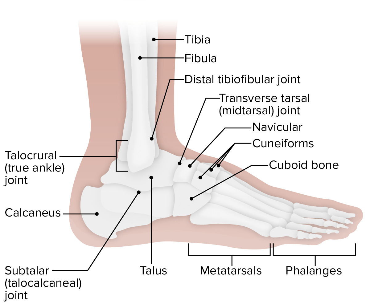
00:01 Okay, let's continue now very distally within the lower limb and talk about the bones of the foot. 00:08 There's a lot of bones here. 00:09 So let's first of all, start by orientating ourselves. 00:12 So we're looking at the superior surface or the dorsal surface of the right foot here. 00:19 And most approximately, we have a series of bones which are known as our tarsals. 00:24 These will then give rise to our metatarsals. 00:27 And then we have some phalanges most distally. 00:30 And these are very similar to the makeup in the hand where we have phalanges most distally, instead of metatarsals, we have metacarpals. 00:37 And instead of tassels, we have carpal bones. 00:40 So the arrangement is very similar in naming. 00:44 But obviously, there's different number and different size and structure of this bony makeup. 00:50 So let's start off by looking at the tarsal group. 00:53 So we've got the tarsal bones here most proximately within the foot. 00:57 The most prominent massive large bone is the calcaneus bone, or you can see it here. 01:03 And then sitting on top of that we have the talus, we then have this clustered as what's known as the proximal group. 01:10 So these two large bones forming the kind of proximal group of this tarsal arrangement of bones within the foot. 01:20 So we see two massive bones here relative to the other ones, the talus, and the calcaneus. 01:25 If we then move anteriorly, we have the navicular bone. 01:28 This is a single bone that forms the more kind of intermediate group of bones within this tarsal group. 01:34 And then finally, we have the cuboid. 01:36 And then three, what are known as cuneiform bones, and these form the distal group. 01:41 So we have three broad groups of bones that form this tarsal arrangement of bones within the foot. 01:49 The proximal group, talus, calcaneus. 01:52 Intermediate group, which is navicular, and then the cuboid and then the three cuneiforms forming the distal group. 01:59 So three groups of bones within this tarsal arrangement. 02:03 If we don't have a look at these from the medial aspects, we can see some of these bones. 02:08 So let's make that transition again, we can see at the top, we've got the talus, and then posteriorly and inferiorly, we've got the calcaneus. 02:17 So let's have a look at the talus bone, which is at the top of the screen there. 02:20 And we hold that in position here. 02:22 Here we can see the talus, we can see the talus as a head, which will go into articulate with the navicular bone, the narrowing neck before it gives rise to the body. 02:32 This body we can call the trochlea. 02:34 And then most inferiorly and working away from the body, we have this posterior process of the talus. 02:42 And these are important landmarks as they help to articulate with the tibia. 02:48 The trochlea specifically helps to run alongside that medial malleolus of the tibia. 02:53 We'll come back to that in a moment or two. 02:56 If we then look more anteriorly, we see how the head of the talus has this articular surface and that articulates with the navicular. 03:05 And then if we add in the calcaneus, we can see the posterior calcaneal surface is articulating with that superior surface of the calcaneus bone. 03:15 So they're essentially we have the talus, which is forming parts of the ankle joint with the tibia and the fibula, we can't see. 03:22 Then inferiorly, we have the calcaneus and then anteriorly, we have the navicular and that's forming part of this medial surface. 03:31 So now if we add on the distal end of the tibia, you can see the medial malleolus is articulating with that articular surface on the superior aspect of the talus and you can see now that inferior aspect of the medial malleolus is articulating with the trochlea that I mentioned a moment ago. 03:48 Here, we're gonna see the articular surface for the distal end of the tibia. 03:51 And you can see the distal end of the tibia highlighted there. 03:55 So we can start to build up having the talus is that central point, anteriorly, the navicular, inferiorly, the calcaneus and then adding on the distal end of the tibia to form the ankle joint. 04:07 So now if we have a look at the lateral view of these bones, we can orientate ourselves again inferiorly, we have the calcaneus. 04:13 And then we have the talus and the navicular and the tibia we can see there. 04:18 As we bring in the fibula, we can see the lateral malleolus. 04:21 And it's articulating on its articular surface with the talus there and again, you can see the anterior calcaneal surface as well. 04:29 If we then bring in this final little region, just a small region, the sulcus tali and that helps to transmit some ligaments, which holds together these bones. 04:38 If we then go back to the medial view, inferiorly, again, we can see the calcaneus, we can see the talus and then anteriorly, we've got the navicular. 04:46 But this time if we have a look at the calcaneus. 04:48 In more detail, we see we have this very broad posterior position calcaneal tuberosity. 04:55 And that's important as it receives the achilles or the calcaneal tendon coming from soleus and gastrocnemius and some other muscles, which we'll come to later on. 05:03 But we've got the broad calcaneal tuberosity sitting there. 05:08 We also have this very kind of like shelf-like structure which is the sustentaculum tali and that little protuberance really allows a pathway for some nerves and arteries and tendons to run underneath as they pass from the leg down on to the sole of the foot. 05:23 And here we can see a groove for the tendon that forms that arrangement, the groove for the tendon of flexor hallucis longus which is going all the way to the big toe first digit. 05:34 Anteriorly the calcaneus, it's going to articulate with the cuboid bone and we can see the articular surfaces there of the calcaneus with the cuboid bone, and then superiorly, we can position the talus on top of it. 05:47 And again, you've got the articular surface there. 05:50 If we then have a look at the lateral view of the calcaneus. 05:53 Again, we can see various structures. 05:55 Here we've got a little groove again, which is for the fibularis longus tendon, and that's got a little shelf above it which helps to create this groove and that's the fibula trochlea. 06:05 So a little elevation on that boat which creates this groove underneath for a tendon to go on. 06:10 We've also got the calcaneal sulcus there and the sulcus tali which we spoke about previously. 06:15 And that's an important region for various ligaments, to run between these bones to hold them in place. 06:21 These together form the sinus tarsi, a little depression that allows various ligaments to move across. 06:29 If we then have a look at the superior view of the calcaneus, we can see how we have various articular surfaces. 06:35 So the anterior tali articular surface, the middle and the posterior one. 06:40 And then between these, we have the calcaneal sulcus. 06:43 Again, this little separation between these various articular surfaces. 06:48 Here we can see the sustentaculum tali which is moving outwards creating that shelf for areas ligaments to various tendons to pass underneath like flexor as long as hallucis that we spoke about previously. 07:00 And here anterior to the calcaneal tuberosity, we have the calcaneal body which we can see located there. 07:07 And again, there is the tuberosity. 07:10 If then we quickly have a look at this from an inferior view or really a posterior view, we can see the calcaneal tuberosity is there, we've got the calcaneal tubercle running superiorly above it. 07:21 And here we have the sustentaculum tali that grew for the tendon of flexor hallucis longus muscle which we have spoken about. 07:28 On the more medial surface, we have a similar arrangement. 07:31 This time it's the fibula trochlea which allows pathway for the fibularis longus tendon to run towards the sole of the foot. 07:38 We'll come back to these when we look at the muscles that are originating from the leg and pass to the foot. 07:44 Now let's have a look at the metatarsals. 07:46 We're nearly there on our journey through the bones of the lower limb and specifically the foot. 07:50 So a few months ago, we've got the metatarsals here, we've got five metatarsal similar to the metacarpals in the bone. 07:58 And for medial to lateral, we have one through to five of these metatarsals. 08:03 Each one of them has a base, a shaft and a head. 08:07 And the head is then going to articulate with the proximal phalanx which we can see there. 08:13 If we move on to these phalanges, we can then see that similar to the hand, the first digit in the foot only has two whereas 2, 3, 4 and 5 has three phalanges. 08:24 So we can see each of them has a proximal phalange there and then we've got a series of middle phalanges and then we've got a series of distal phalanges there. 08:33 So we can see that we've got two making up the first digit for the big toe and then three making up all of the subsequent digits from two through to five. 08:45 Each of these phalanges similar to the metatarsals, has a base, a shaft and a head. 08:51 And then these together can form a series of joints across the entire foot working all the way from the posterior aspects of the calcaneus aspects all the way through to the inter phalangeal joints we see most distally and we can talk about those in a moment or two when we look at the movement. 09:09 But specifically, if we go back to the phalanges, which we're talking about, you got the inter phalangeal joints between the phalanges and the metatarsals, you have the metatarsal phalangeal joints. 09:19 And then between the tarsal bones and the metatarsals, you have the tarsometatarsal joint and then you have various joints between the tarsal bones. 09:28 So the subtalar joint inferior to the talus bone articulating with the calcaneus. 09:35 You also have the transverse tarsal joint that's running across these tarsal bones, holding them all into position. 09:41 So you can see a whole number of joints that are articulated there within this lateral view of the foot.
About the Lecture
The lecture Bones of the Foot by James Pickering, PhD is from the course Osteology and Surface Anatomy of the Lower Limbs.
Included Quiz Questions
Which foot bone(s) is/are most distal?
- Phalanges
- Metatarsals
- Calcaneus
- Talus
- Navicular
There are how many cuneiform bones in each foot?
- 3
- 2
- 4
- 5
- 7
Which bones are considered the distal group of the tarsal bones? Select all that apply.
- Medial cuneiform
- Cuboid
- Navicular
- Talus
- Calcaneus
Which bone articulates with the tibia?
- Talus
- Navicular
- Calcaneus
- Medial cuneiform
- Lateral cuneiform
Where does the Achilles tendon attach?
- Calcaneal tuberosity
- Articular surface for navicular
- Posterior calcaneal surface
- Medial malleolus
- Lateral malleolus
There are how many metatarsal bones in each foot?
- 5
- 4
- 3
- 2
- 1
Which movements are possible at the hip joint? Select all that apply.
- Flexion
- External rotation
- Internal rotation
- Abduction
- Dorsiflexion
Author of lecture Bones of the Foot

James Pickering, PhD
Customer reviews
5,0 of 5 stars
| 5 Stars |
|
5 |
| 4 Stars |
|
0 |
| 3 Stars |
|
0 |
| 2 Stars |
|
0 |
| 1 Star |
|
0 |
