Playlist
Show Playlist
Hide Playlist
Anterior View of the Brain Stem

-
Slides 6 BrainStem BrainAndNervousSystem.pdf
-
Download Lecture Overview
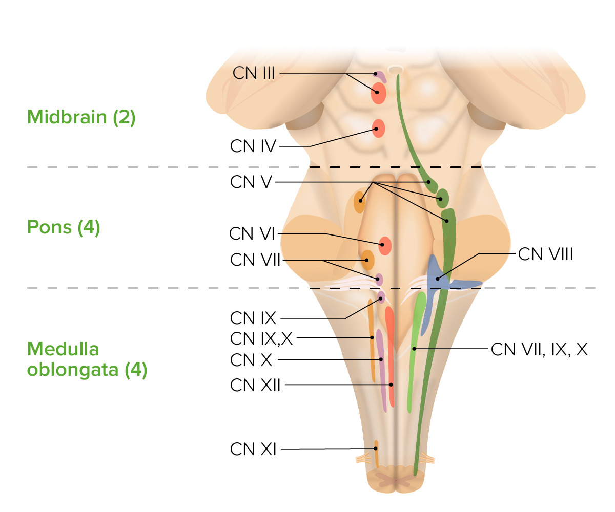
00:00 Here is an anterior view of the brainstem region. This represents or demonstrates the cranial nerves in this anterior view. This is just a quick listing of them. You can see cranial nerve number III. 00:22 You see cranial nerve number IV coming from the posterior aspect of the brainstem and coursing around the pons area. Here’s cranial nerve number V. Here’s the abducens nerve, cranial nerve number VI. 00:41 Here’s VII, here’s VIII, number IX, number X. Here is XI, the accessory. Then lastly, you have cranial nerve number XII. This is just a quick summary of the relative positions of the cranial nerves from III through XII in an anterior view. We’re going to maintain this anterior view. What I want you to realize are the structures that can be seen here and the functions that are attached to these structures. 01:23 In this particular view right in through here, superior to the pons, you can see some prominent structures. 01:30 Each one represents the crus cerebri. This is a major pathway of motor output from the cortex. 01:40 These descending fibers are going inferiorly through the brainstem, travel through the brainstem, and then will continue their journey into the corticospinal tracts of the spinal cord. The pons, we see right in through here, very prominent brainstem structure. Again, cranial nerve nuclei V through VIII are found here. Corticospinal fibers are descending through here. Corticobulbar motor fibers are also descending through the pons. We have fibers between the pons and the cerebellum travelling in the pons, pontocerebellar fibers. The medial lemniscus is found in this structure. Medial longitudinal fasciculus is another structure found here. The pontine center for lateral gaze and the reticular formation is also found here and is found elsewhere within the brainstem. Now, we’re going to look at the medullary area. We have a very prominent dilatation on the lateral aspects called the olive. 03:01 It’s hidden from view here by the hypoglossal nerve on the opposite side. With the cranial nerve out of the way, you can see this dilatation. The olive is involved in cerebellar motor learning. 03:15 Then, more centrally located is the medullary pyramid. We see the opposite one over here. The pyramids represent descending motor tracts, specifically, these are the corticospinal tracts descending within the medullary pyramids. Most of the fibers that make up these corticospinal tracts are decussating or crossing in through here. 90% of those fibers do cross or decussate. The other 10% will remain ipsilaterally and continue their descending path.
About the Lecture
The lecture Anterior View of the Brain Stem by Craig Canby, PhD is from the course Brain Stem.
Included Quiz Questions
Which of the following is NOT a feature of the pons?
- Oculomotor nerve nuclei
- Corticospinal motor fibers
- Pontocerebellar fibers
- Medial longitudinal fasciculus
- Reticular formation
Which of the following is involved in cerebellar motor learning?
- Inferior olivary nucleus
- Medullary pyramid
- Pretectal nucleus
- Medial longitudinal fasciculus
- Reticular formation
Author of lecture Anterior View of the Brain Stem

Craig Canby, PhD
Customer reviews
5,0 of 5 stars
| 5 Stars |
|
5 |
| 4 Stars |
|
0 |
| 3 Stars |
|
0 |
| 2 Stars |
|
0 |
| 1 Star |
|
0 |
