Playlist
Show Playlist
Hide Playlist
Anterior Compartment of the Thigh
-
Slide Anterior Compartment of the Thigh.pdf
-
Download Lecture Overview
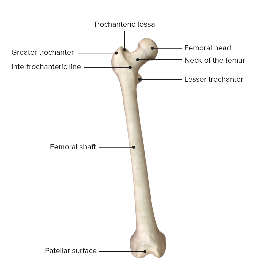
00:01 Now let's spend the time looking at the anterior thigh muscles. 00:05 So these muscles broadly are important in extending the leg at the knee joint, they can also help as some of them cross from the pelvis and the vertebral column into the thigh. 00:17 They can also help with flexion of the femur at the hip joint, but primarily the anterior thigh muscles are we going to look at are responsible for extending the leg at the knee joint. 00:28 Let's go through some of the muscles that we'll talk about. 00:30 We have iliopsoas muscle here, we have sartorius. 00:34 We also have quadriceps femoris muscle here. 00:38 And if we look at these in slightly more detail, here we have iliopsoas, we have psoas major, which is coming down from the lateral aspect of the vertebral column, and it's united with iliacus muscle from the internal surface of the ilium. 00:52 These two muscles unite together to form iliopsoas. 00:56 The origin of psoas muscle comes from, like I said, the lateral surface of the vertebrae, specifically T12 through two L5. 01:06 Iliacus muscle is coming from the iliac crest and the fossa of the ilium. 01:10 And they pass down and attached to the lesser trochanter of the femur. 01:15 They're important because they run anterior to the hip joint and attached to the femur. 01:20 They're important in flexion of the thigh at the hip joint. 01:25 Next we have the sartorius muscle which is very long, slender muscle that has quite a unique range of movement due to the way it's orientated within the lower limb. 01:35 Here we can see sartorius originating from the anterior superior iliac spine of the ilium. 01:43 And it runs all the way down to attach to the tibial tuberosity. 01:47 This muscle though, takes quite an interesting course. 01:51 And as it does, so it means it has a wide range of movement. 01:54 So because there is broad range of positioning within the thigh region, and it crosses anteriorly to the hip joint, it's responsible for flexing, abducting and laterally rotating the hip. 02:06 It also is responsible for extending the knee joint. 02:09 So sartorius has a broad range of functions. 02:13 Now let's look at quadriceps femoris. 02:15 This is an interesting muscle because it's really made up of four different muscle bellies. 02:20 We've got rectus femoris, vastus medialis, vastus lateralis, and if we remove rectus femoris, we see deep to it vastus intermedius. 02:31 These muscles then converge on the quadriceps tendon, which then runs over the patella to form the patellar ligament and attaches onto the tibial tuberosity. 02:41 If we go through them from deep to superficial, vastus intermedius comes from the anterior side of the femoral shaft, vastus lateralis comes from the lateral surface of the greater trochanter and the lateral lip of the linear aspera. 02:56 You may want to go back and look at the bony features we looked at the femur in a previous video. 03:02 Vastus medialis coming for the more medial aspect comes from the intertrochanteric line and the medial lip of the linear aspera. 03:11 And then rectus femoris is coming from the anterior superior iliac spine. 03:16 Notice how this aspect of quadratus femoris is actually crossing the hip joint because it's coming from the ilium. 03:24 So rectus femoris comes from the anterior superior iliac spine, and all of these muscles converge via the quadriceps tendon over the patella to the patellar tendon and then on to the tibial tuberosity. 03:38 These muscles are associated with flexion of the hip because of the movement of rectus femoris. 03:43 And they're also associated with extension of the knee joint because they run anterior to the knee joint. 03:49 So they extend the leg at the knee joint. 03:53 Now let's have a look at the muscles of the anterior compartment and their innovation. 03:58 So here, we can see psoas as major is innovated by the anterior rami of L1, L2, L3. 04:04 We can then see iliacus, quadriceps femoris and sartorius are all innovated by the femoral nerve. 04:11 Remember the femoral nerve is coming L2, L4.
About the Lecture
The lecture Anterior Compartment of the Thigh by James Pickering, PhD is from the course Anatomy of the Thigh.
Included Quiz Questions
What is the primary function of the anterior thigh muscles?
- Extend the leg
- Flex the leg
- Abduct the leg
- Adduct the leg
- Internally rotate the leg
What is the origin of the psoas major muscle?
- T12 - L5 vertebrae
- T10 - L5 vertebrae
- T10 - L2 vertebrae
- T10 - L3 vertebrae
- T12 - L3 vertebrae
What is the origin of the sartorius muscle?
- Anterior superior iliac spine
- Posterior superior iliac spine
- Tibial tuberosity
- Fibula tuberosity
- Iliac crest
Customer reviews
5,0 of 5 stars
| 5 Stars |
|
5 |
| 4 Stars |
|
0 |
| 3 Stars |
|
0 |
| 2 Stars |
|
0 |
| 1 Star |
|
0 |


