Playlist
Show Playlist
Hide Playlist
Anterior Compartment of the Leg
-
Slide Anterior Compartment of the Leg.pdf
-
Download Lecture Overview
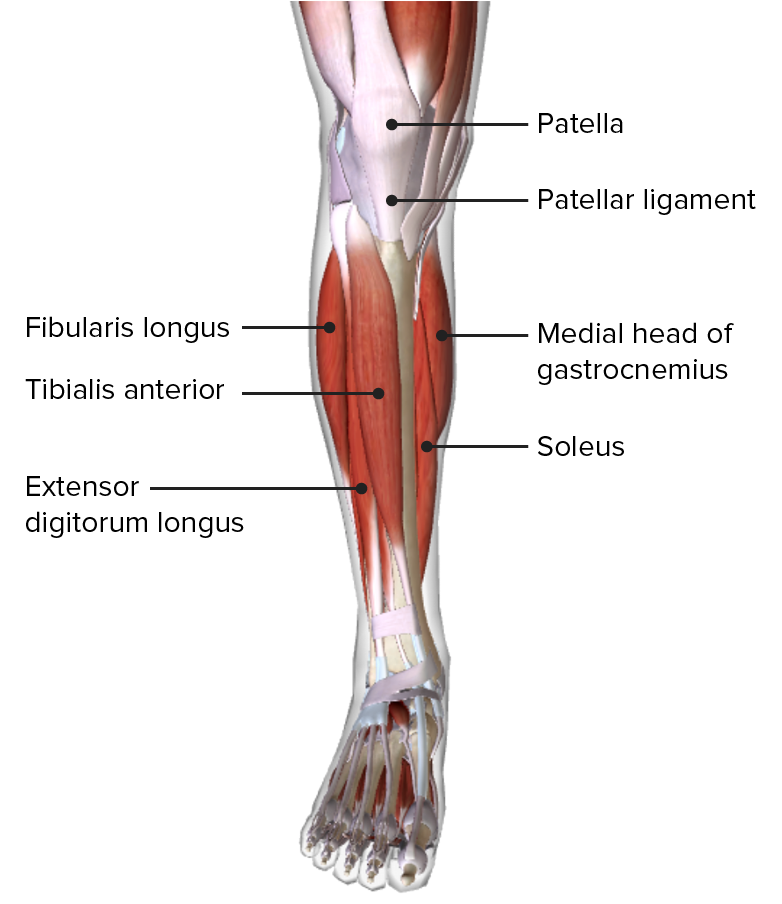
00:01 So let's have a look at the muscles within this anterior compartment of the leg. 00:06 And to orientate ourselves, we're looking at the anterior surface of the leg within the right lower limbs. 00:11 So we can see the tibia medially and the fibular laterally. 00:15 Here we can start adding in some of these muscles, we find in this anterior compartment. 00:21 We have a very small muscle which is known as fibularis tertius, we can see here. 00:25 Then we have a longer muscle, extensor digitorum longus, we can see here. 00:30 We then have extensor hallucis longus, and we have tibialis anterior. 00:36 Remember some of these names will help us to identify what these muscles do. 00:40 So extensor means it's going to extend, digitorum means it'll extend the digits. 00:45 And the longus version means later on, we'll have a brevis version. 00:49 So we'll have extensor digitorum longus. 00:52 And later on, we'll realize we have extensor digitorum brevis. 00:55 So two muscles responsible for extending the digits, so you can use a terminology to help you understand what these muscles do and where they go. 01:05 Let's have a look at some of the origins, insertions and actions of these muscles, starting with tibialis anterior. 01:12 We can see tibialis anterior here, it kind of travels down in an inframedial position. 01:18 It originates from the lateral condyle of the tibia, the lateral surface of the proximal tibia, and also the interosseous membrane. 01:28 It runs down onto the medial surface of the foot, where it then attaches to the medial cuneiform and the base of the first metatarsal. 01:37 It has a couple of movements, it helps to dorsiflex the ankle, and it also helps to invert the foot. 01:45 Now let's have a look at extensor digitorum longus. 01:49 This muscles runs very straight down alongside the fibular. 01:53 It originates from the lateral condyle of the tibia. 01:56 It also has attachments on the anterior surface of the fibular, and the interosseous membrane. 02:02 It runs down and passes towards the middle and distal phalanges of the second to fifth digits. 02:09 So now let's turn to the function of extensor digitorum longus. 02:13 It helps to dorsiflex the ankle and it also helps to extend the second to fifth digits. 02:20 Now let's move on to extensor hallucis longus. 02:23 Here we can see extensor hallucis longus is originating from the interosseous membrane and the anterior surface of the mid fibular point. 02:32 It passes all the way down to the distal phalanx of the first digit, and as its names applies, hallucis indicates the great toe, big toe, the first digits of your foot. 02:44 The movement of this muscle is too fold again, it's dorsiflexion of the ankle and also it helps to extend the first digit. 02:53 If we then move on to the final muscle within this compartment, we have fibularis tertius. 02:57 This is quite a small muscle on the anterior inferior aspect of the leg. 03:02 It originates from the inferior portion of the interosseous membrane, and also the anterior surface of the lower fibular around the lower third. 03:11 It passes all the way down to the fifth metatarsal. 03:15 The function of this muscle is again to support dorsiflexion. 03:19 It also helps to support a version of the foot when you're elevating the little toe. 03:25 Now let's look at the innervation to the anterior compartment of muscles. 03:30 This is primarily done via the deep fibular nerve. 03:34 And if we can remind ourselves, the deep fibular nerve is a branch from the tibial from the sciatic nerve when it branches into the common fibular and the tibial. 03:43 We'll have a look at that again in a moment. 03:46 But what we can see here is the deep fibular nerve passing deep to an extensor retinaculum. 03:51 As it passes towards the foot, this inferior extensor retinaculum is Y-shaped and it attaches medially to both the calcaneus and the medial malleolus. 04:03 Its lateral attachment is at the calcaneus on the lateral aspect. 04:07 There's also a superior extensor retinaculum and this these together help to support the tendons within the correct position to prevent them from both stringing during flexion and plantar extension dorsal extension of the foot and ankle. 04:22 You can see the fibular is most lateral in this position. 04:24 And importantly, the superior extensor retinaculum is then just running towards the fibular as it crosses over from the tibia.
About the Lecture
The lecture Anterior Compartment of the Leg by James Pickering, PhD is from the course Anatomy of the Leg.
Included Quiz Questions
What is the origin of the tibialis anterior? Select all that apply.
- Lateral condyle
- Interosseous membrane
- Medial cuneiform
- Medial metatarsals
- Middle and distal phalanges
What is the insertion site of the extensor digitorum longus?
- Middle and distal phalanges
- Lateral condyle
- Anterior surface of fibula
- Interosseous membrane
- Posterior surface of fibula
Customer reviews
5,0 of 5 stars
| 5 Stars |
|
5 |
| 4 Stars |
|
0 |
| 3 Stars |
|
0 |
| 2 Stars |
|
0 |
| 1 Star |
|
0 |


