Playlist
Show Playlist
Hide Playlist
Ankle Joint

-
Slide Ankle Joint.pdf
-
Download Lecture Overview
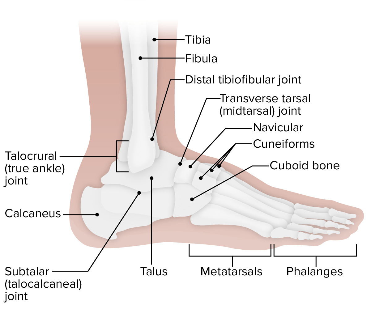
00:01 Now let's move to the ankle joint. 00:03 The ankle joint is a hinged synovial joint. 00:07 So here we can see the ankle joint is articulating between three bones - the fibular, and the tibia superiorly, and the talus inferiorly. 00:16 And here we can see specifically on the talus, we have the trochlea. 00:20 This is rarely wide anteriorly but more posteriorly becomes narrower, and this helps to wedge the ankle joint together to help increase stability. 00:30 We'll come back to that later on. 00:32 But the trochlea of the talus is very important in that it's quite wide anterior relatively to a narrow posterior formation of the talus. 00:43 Here we have the malleolar mortise. 00:45 And this is the aspect that sits on that talus. 00:48 So you can appreciate that as the ankle moves, and the wider anterior portion moves into the malleolar mortise. 00:56 As its wider, it increases stability. 01:00 And then, for movement of the ankle to occur, you need that posteriorly narrow portion to move into the malleolar mortise that increasing the space for you to move. 01:10 So this wedge formation, as it indicated earlier, helps to increase the stability of this ankle joint. 01:18 The lateral wall here on the fibular is the lateral malleolus of the fibular. 01:23 Then you have a roof of this mortise, which is the inferior surface of the distal end of the tibia. 01:29 And then on the medial wall, you have that medial malleolus of the tibia, you can see there. 01:34 You have a little recess, the lateral part of the trochlea, which just lies on the lateral aspect of the talus, and that is where the lateral malleolus of the fibular can rest. 01:44 Here we see the two articular surfaces, the inferior surface of the tibia, and the central part of the trochlea. 01:50 And these two will come together at the ankle joint. 01:54 The medial part of the trochlea is that piece that's going to side against the medial malleolus of the tibia. 01:59 And here we have our articular surfaces of the ankle joint. 02:04 The capsule is running all the way around the ankle joint. 02:08 So it's surrounding both the fibular, the tibia, and the talus. 02:12 And here we're gonna see the anterior margin of the tibia and fibular. 02:15 And here we see the articular margins around the talus. 02:19 So these are the extremes of the joint capsule. 02:22 So now let's have a look at the ligaments that helped to support the ankle joint. 02:26 These are collectively known on the medial side as the medial ankle joint ligaments, or the deltoid ligaments. 02:33 There's a number of these that helped to stabilize this medial aspect of the ankle joint. 02:38 Here we can see the anterior tibioalar part of this deltoid joint. 02:43 It's coming from the medial malleolus and passes all the way down to the medial tubercle of the talus. 02:49 Here we have the tibionavicular part. 02:51 This is running from the medial malleolus all the way down to the navicular. 02:56 And then we have the tibiocalcaneal parts, again running from the medial malleolus, and this time passing to the sustentaculum tali of the calcaneus. 03:05 We have the posterior tibioalar apart, and that's running from the medial malleolus of the tibia, all the way down to the medial tubercle of the talus. 03:14 So we have a number of parts that constitute this deltoid ligament of the ankle joint. 03:20 The anterior posterior tibioalar, then the tibionavicular, and tibiocalcaneal. 03:26 These ligaments are essentially named after the bones they attach to. 03:30 If we then look on the lateral aspect, we find the lateral ligaments of the ankle. 03:35 Again, helping to reinforce and stabilize the ankle joint around the joint capsule. 03:40 This time we have an anterior talofibular ligament that's running from the lateral malleolus of the fibular this time. 03:47 And that passes to the adjacent region of the talus. 03:51 We also have a calcaneofibular ligament running from the lateral malleolus of the fibular to the calcaneus' lateral surface. 04:00 As we add an anterior talofibular ligament, we also have a posterior talofibular ligament, again running from the lateral malleolus of the fibular down to the posterior process of the talus. 04:12 So we can see we have three ligaments that make up this lateral ligament of the ankle. 04:18 On the medial aspect, we had four. 04:20 But this time we have three on this lateral aspect - anterior and posterior talofibular, and the calcaneofibular ligament as well. 04:29 These ligaments both lateral and medial are helping to reinforce the joint capsule and stabilize the ankle joint. 04:36 The vascular supply to the joint capsule around the ankle is by way of the anterior and posterior tibial arteries, which we're familiar with as we've looked at the arterial supply of the leg. 04:49 These are coming down from the popliteal artery. 04:51 We also have contributions from the fibular artery and these give rise to branches around the medial and lateral malleolus. 04:58 So we can see these lateral and medial malleolar arteries helping to supply the joint capsule. 05:05 Let's have a look at the movements that can occur at the ankle joint. 05:08 So here we have a dorsiflexion where the foot is being lifted up off the ground. 05:14 And here we can see the opposite of dorsiflexion at that hinge of the ankle joint is plantarflexion, when you're moving to stand on your tiptoes.
About the Lecture
The lecture Ankle Joint by James Pickering, PhD is from the course Joints of the Lower Limbs.
Included Quiz Questions
Which ligament does NOT support the medial ankle joint?
- Anterior talofibular
- Anterior tibiotalar
- Tibionavicular
- Tibiocalcaneal
- Posterior tibiotalar
Which ligament supports the lateral part of the ankle joint?
- Calcaneofibular ligament
- Anterior tibiotalar ligament
- Tibionavicular ligament
- Tibiocalcaneal ligament
Which arteries provide blood to the ankle joint? Select all that apply.
- Anterior tibial artery
- Lateral malleolar artery
- Medial tibial artery
- Lateral tibial artery
- Posterior malleolar artery
Author of lecture Ankle Joint

James Pickering, PhD
Customer reviews
5,0 of 5 stars
| 5 Stars |
|
5 |
| 4 Stars |
|
0 |
| 3 Stars |
|
0 |
| 2 Stars |
|
0 |
| 1 Star |
|
0 |
