Playlist
Show Playlist
Hide Playlist
Angiogenesis in Wound Healing

-
Slides Acute and Chronic Inflammation Angiogenesis.pdf
-
Download Lecture Overview
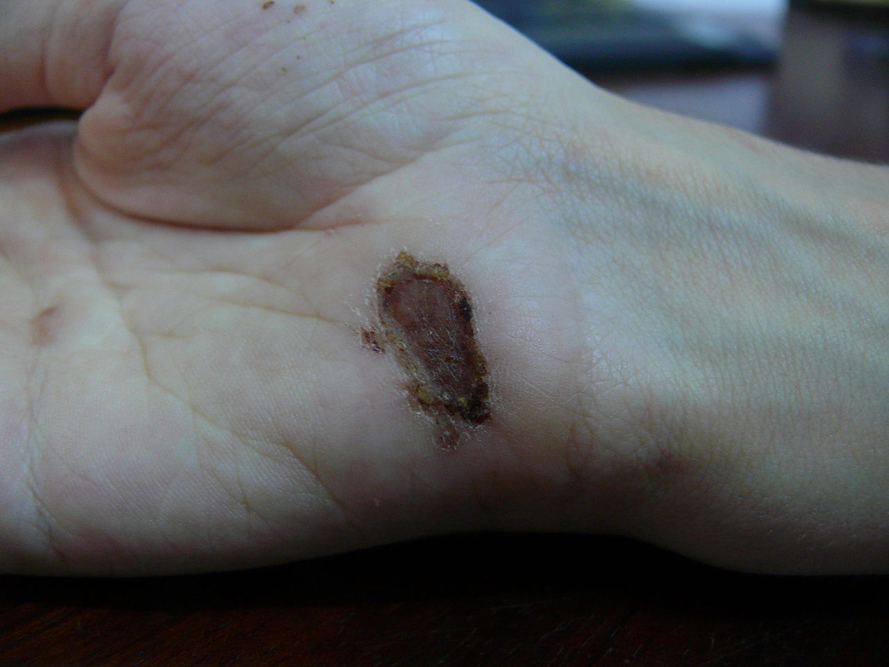
00:01 All right, then. 00:02 Let's move along. 00:03 We have moderated acute inflammation. 00:07 We're into the stages now, where we are really trying to get the definitive healing to go on. 00:15 In this process when we cannot get regeneration, we need to lay down scar. 00:21 And the first step in that is angiogenesis. 00:25 Here's a roadmap. 00:26 You can see we've already covered the top three topics, moderating acute inflammation, recruiting and activating macrophages, regenerating parenchyma, if we can. 00:37 The next two are what about not being able to regenerate and that starts as he said with angiogenesis. 00:45 Angiogenesis is recognized by us as pathologists as granulation tissue. 00:54 A word granulation tissue is not a granuloma. 00:59 Remember, granulomas are a nodule of activated macrophages. 01:03 Granulation tissue is this early provisional stroma with a lots of new blood vessels upon which we're going to lay down scar. 01:13 So granulation tissue is no more like a granuloma than it is like your grandma. 01:19 Okay, just make that really clear. 01:23 This is granulation tissue. 01:25 It's going to peak in tissues that have been injured at about five to 10 days. 01:30 It's very loose connective tissue. 01:33 It's got a lot of edema, because the vessels are incredibly leaky. 01:37 It's got a few residual inflammatory cells, but mostly what granulation tissue is, are these small, new, delicate capillaries that we've just built. 01:48 And why is it called a granulation tissue? Well, if you're anything like me, I cannot leave a scab alone. 01:53 So when I have a scab, I pick it. 01:55 And underneath are these little pink granules, they kind of they leak blood, because there was a very delicate capillaries. 02:02 And the surgeons of your recognize that in a wound that had lots of good granulation tissue, that wound was going to do well, it was going to heal in. 02:11 But if there wasn't good granulation tissue, because of the little, you know, the capillaries weren't forming, well, that was going to likely get infected. 02:19 So it's called granulation, because the little apparent granules, a pink capillaries that you can see under a scab. 02:25 Okay. That's a sidelight. 02:28 In any event, this is what it looks like. 02:31 What it is? New Delicate Vasculature Has very loose extracellular matrix, as you can see there. 02:38 There are residual chronic inflammatory cells. 02:40 In fact, the macrophages mostly M2 macrophages at this point, are going to be responsible for coordinating all the next few steps. 02:49 And in fact, they're the major source driving the formation of the granulation tissue. 02:53 So they need to be there to do this. 02:56 It's very edematous, because the vessels are leaky. 03:00 And it bleeds very easily. 03:02 As what happens when you pick that scab and the little vessels bleed. 03:06 It's the scaffolding. 03:07 It's the provisional matrix upon which we're going to lay down scar. 03:11 Okay, so that's, it's all of those things. 03:14 So how exactly do we get these new blood vessels? What is the mechanism of angiogenesis? All right, on the right hand side of your screen, it's a big white area. 03:25 That's a dead zone, that is where we've had tissue injury and all kinds of death and destruction occur there. 03:31 There will be macrophages that are out there, because they're cleaned up the debris, and they're going to be the ones driving the next stages. 03:37 So the very first stage of angiogenesis, we have to break down the membrane, the basement membrane, at the nearest blood vessel that's intact. 03:49 So we need to be able to have a sprout of new blood vessels that will grow into the dead zone. 03:54 And that proteolysis of extracellular matrix is driven by molecules, mediators, elaborated by the macrophages. 04:01 So you see the macrophages there, and those little dots, those are going to be various factors that are going to drive the breakdown of extracellular matrix. 04:10 What are those? Some of the factors there are others, but the two main ones are basic FGF, basic fibroblasts derived growth factor made by macrophages. 04:23 And then you have vascular endothelial growth factor or VEGF, also made by macrophages and other cells. 04:29 But that's going to start the first process of breaking down the basement membrane, and allowing us to have a bud of new capillary growth. 04:38 Next step, we need to have the endothelial cells that are there at that new bud migrate in the direction of where we want new blood vessels to form. 04:48 So we want them to migrate to chemotaxis. 04:51 And you see the macrophages out there spewing some more little bubbles, and those are going to be the factors that drive this process. 04:59 And what are the those? Wow, it's basic FGF and VEGF again. 05:05 So far, we only have to memorize two additional factors, for the most part. 05:11 Basic FGF and VEGF, cause the proteolysis of the bsal membrane and also now drive the chemotaxis of the endothelial cells out into the dead zone. 05:21 Okay, well, it's just not enough to have them migrate. 05:26 Clearly, they can't get stretched thinner, and thinner, and thinner. 05:28 We need to make more of them in order to make a blood vessel of new capillary. 05:32 So they have to proliferate. 05:34 And again, you see the macrophages out there and the dead zone spewing pixie dust to make this happen. 05:40 What's in the pixie dust this time? Wow, same stuff, basic FGF and VEGF, and that's going to be driving proliferation of the endothelial cells. 05:50 Cool. 05:51 Okay, it's not just enough that we have a whole bunch of endothelial cells growing and marching into the dead zone. 05:57 They need to form a tube so that we can bring in blood. 06:00 So we need to actually form a mature capillary. 06:05 That maturation has to also happen with kind of an inhibition of growth. 06:09 We want things to stop. 06:11 How do we make that happen? The lumen formation, the maturation, and inhibition of growth, Do you think it's basic of FGF and VEGF? No, unfortunately, it's not. 06:21 It's two other factors, or it's one other factor called angiopoietin-1 that's elaborated by perisites, P-E-R-I sites that are part of the maturing capillary. 06:34 And there are receptors for angiopoietin-1 called Tie-2 that live on the endothelial cells. 06:41 So that maturation process, different set of factors, unfortunately. 06:47 In the process of angiogenesis, we've got a mature blood vessel that's out there, but we are now going to have a very highly metabolically active tissue. 06:55 We're going to lay down a lot of new tissue out there. 06:58 We need a fair amount of nutrition to make that work. 07:02 So one of the ways that we can improve that deposition of nutrition is to make the vessels a little extra leaky, so that more nutrition gets out fairly easily, without having to be transported. 07:15 To do that, we need to increase permeability through gaps or through transcytosis moving contents that are in the lumen out into the newly forming scar tissue. 07:29 Again, macrophages are driving this with little bits of of pixie dust, a little balls, and what's in there? Again, basic FGF and VEGF. 07:40 So for most of the factors involved in angiogenesis, we can actually blame it, or attributed to basic FGF and VEGF.
About the Lecture
The lecture Angiogenesis in Wound Healing by Richard Mitchell, MD, PhD is from the course Acute and Chronic Inflammation.
Included Quiz Questions
Which of the following histological features is associated with granulation tissue?
- Formation of new thin-walled capillaries
- Accumulation of macrophages and necrotic debris
- Extensive interstitial collagen deposition
- Inflammatory infiltrate with abundant neutrophils
- Interstitial deposition of lipofuscin pigment granules
Which of the following is involved in capillary maturation?
- Angiopoietin-1
- Vascular endothelial growth factor
- Basic fibroblast growth factor
- Tissue necrosis factor-alpha
- Epidermal growth factor
Which of the following is true about vascular endothelial growth factor (VEGF)?
- It promotes the proteolysis of the extracellular matrix.
- It is produced and secreted by the pericytes.
- It is involved in the inhibition of endothelial growth and lumen formation.
- It binds to the Tie-2 receptor on the endothelial cells.
- It drives the chemotaxis of neutrophils and red blood cells.
Which of the following increases the vascular permeability during angiogenesis?
- Basic fibroblast growth factor
- Angiotensin
- Thromboxane A2
- Epidermal growth factor
- Interleukin 10
Author of lecture Angiogenesis in Wound Healing

Richard Mitchell, MD, PhD
Customer reviews
5,0 of 5 stars
| 5 Stars |
|
5 |
| 4 Stars |
|
0 |
| 3 Stars |
|
0 |
| 2 Stars |
|
0 |
| 1 Star |
|
0 |
