Playlist
Show Playlist
Hide Playlist
Anatomy of the Duodenum

-
Slides Anatomy of the Duodenum.pdf
-
Download Lecture Overview
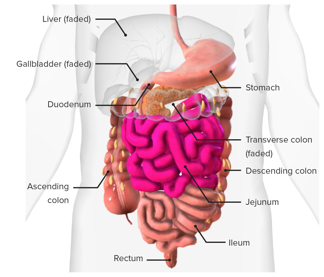
00:01 So let's now have a look in more detail at the duodenum. 00:04 So as we've spoken before, the duodenum is a C-shaped structure or C-shaped extension of the tube that comes from the stomach and it very much follows the contour of the pancreas. We can see the pancreas in the central part of the screen there. So coming away from the stomach, we have the pyloris which gives rise to the first part of the duodenum, the first part of the duodenum also known as the superior part. Here, we have the superior part going towards the right. It then takes a sudden downward trajectory and forms the descending part. So you can see it takes quite an abrupt leftward turn to run down alongside the head of the pancreas before it then takes another sharp turn this time to the left again where it's forming the horizontal part. So here we have the first 3 parts of the duodenum; superior, descending, horizontal and then it ascends up as the ascending part of the duodenum. 01:01 So 4 parts of the duodenum there. The 4th part of the duodenum gives rise to the jejunum and that is at the duodenojejunal junction, a really important part as you start having a transition in the microstructure of the small intestine. Here we can see the important in a surface of the duodenum and how it relates to the structures that are surrounding the duodenum. So as I mentioned previously, there's a large number of accessory organs of digestion that feed in various pancreatic hepatic juices into the tube to aid with digestion. And here we can see the start of this. So here if we have a closer look at the inside wall of the duodenum, we can start to see those circular folds to help again to increase the surface area. But we can begin to see an opening that is coming from both the pancreas and the liver. And this is known as the major duodenal papilla. It's a little opening that is receiving a tube which is coming from the liver. This is the common bile duct. And what's coming from the common bile duct is bile. This helps to breakdown fats. 02:10 Bile is stored in the gallbladder but is produced in the liver. So bile will come down from the liver and head towards the duodenum. If this major duodenal papilla is closed, then bile will back up and it will be stored in the gallbladder. What you also have in this opening is the main pancreatic duct. 02:32 And this is bringing in pancreatic juice from the pancreas, enzymes such as amylase, etc. and that helps to breakdown the food that's being ingested within the gastrointestinal tract. But what we have here is this large opening, this ampulla. And this ampulla is receiving both the common bile duct and the main pancreatic duct and the union of these 2 tubes then pass into the duodenum by the major duodenal papilla. We also, slightly superior to it, have a minor duodenal papilla. This is not seen everywhere and it's predominantly a second reroute for pancreatic juice to leave the pancreas and passing to the duodenum. It doesn't really have any connection with the common bile duct. And here we can see the accessory pancreatic duct feeding in to this major duodenal papilla. You can see the arrow there indicates the movement. So here we can see the duodenum and it's slightly orientated to the left stomach and duodenum. 03:35 Each here we can see the stomach and it's giving rise to the duodenum which you can see there. That's the first part of the duodenum. Here we got the common bile duct that's passing down and passing in thru the substance of the pancreas to then go and enter into the descending parts of the duodenum you can see there. Also running alongside the common bile duct, you can see the gastroduodenal artery. We spoke about that one, we looked at the blood supply to the stomach. And you've also got the hepatic portal vein which is running in this space again. We'll have a look at these structures a lot more when we talk about the peritoneum as they're associated in various ways with the lesser omentum and the free edge of the lesser omentum, and that's an important structure. We'll talk about that in a moment or two. Here we can see the duodenum is associated with right kidney and then as we look towards the bottom of this region, we can also make out the horizontal parts of the duodenum, the third part. Close approximation with the right psoas muscle running along the posterior aspect of the abdominal wall and also the right ureter. You'll be able to trace the right ureter on the screen coming descending down from the kidney. Here, we can see the inferior vena cava as well. And then finally, we have the 4th part of the duodenum which remember is then ascending slightly to form the duodenojejunal junction and here running next the inferior vena cava we have the abdominal aorta. Similar to the right hand side, we also see the close association with the ureter not highlighted but as we just saw there you have the left psoas major muscle as well. There's a lot going on in this region and here looking at the more posterior aspect of the duodenum you do see there's a lot of structures, a lot of associated with this region indicating actually the depth of the duodenum and how it's situated against the posterior abdominal wall alongside the pancreas. The pancreas very much sits in the concavity of the C shape of the duodenum and if you remember previously when we spoke about how superficial the stomach was to the anterior abdominal wall, I hope this does give an indication that actually the pancreas is deep and therefore operating on the pancreas can be very difficult. As you can see, there are lots of neighboring structures. Here, we can see laterally we got the ascending colon and running all the way around the duodenum, obscuring it, we're going to have the jejunum. It does become a very complicated structure if you're wanting to move various organs out of the way. We introduce the gallbladder, we introduce the liver, and you can see actually getting into the duodenum can be a terribly difficult structure to access. The transverse colon is probably one of the first organs especially when you have to move to the greater omentum as well to actually start seeing the duodenum clearly. 06:19 So operating around the pancreas or pancreatic cancer, for example, can be incredibly, incredibly difficult.
About the Lecture
The lecture Anatomy of the Duodenum by James Pickering, PhD is from the course Anatomy of the Small Intestine.
Included Quiz Questions
In which part of the duodenum is the major duodenal papilla located?
- Descending
- Superior
- Horizontal
- Ascending
- Posterior
The major duodenal papilla is located at the opening of which structure that leads into the duodenum?
- Bile duct
- Accessory pancreatic duct
- Hepatic duct
- Cystic duct
- Cystic artery
Which structure sits in the concavity of the duodenum?
- The head of the pancreas
- The tail of the pancreas
- The neck of the pancreas
- The gallbladder
- The spleen
Author of lecture Anatomy of the Duodenum

James Pickering, PhD
Customer reviews
5,0 of 5 stars
| 5 Stars |
|
5 |
| 4 Stars |
|
0 |
| 3 Stars |
|
0 |
| 2 Stars |
|
0 |
| 1 Star |
|
0 |
