Playlist
Show Playlist
Hide Playlist
Anal Canal

-
Slides Anal Canal.pdf
-
Download Lecture Overview
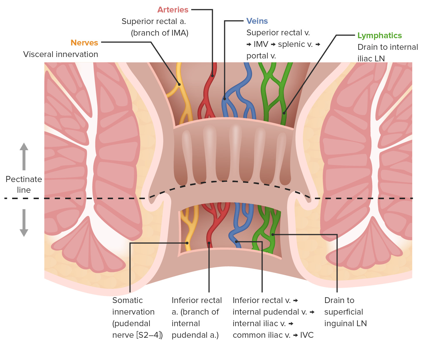
00:01 Now, let's have a look at the anal canal. 00:04 So, the anal canal is residing within the anal triangle of the perineum. 00:09 So, here we can see some familiar landmarks to help orientate ourselves. 00:13 So, what we've done is we've cut through a coronal section through the pelvis, specifically the level of the anus. 00:20 So, here we can see the ischium. 00:22 Here we can see obturator internus, forming that lateral wall. 00:26 Here we can see levator ani, forming that angled medial wall. 00:30 And here we can see the fatfield space of the ischioanal fossa. 00:35 Notice again, that there isn't a penny or membrane here, separating this space into deep and superficial layers. 00:43 We've just got a completely wedge shaped ischio-anal fossa, which is filled with fat. 00:50 Here we can see above levator ani muscle is the pelvic cavity. 00:54 So now, we can appreciate how the rectum becomes the anal canal in the anus as it passes through the anal aperture of the pelvic diaphragm to then reside within the anal triangle of the perineum. 01:07 So, here we can see the pelvic cavity. 01:09 Here we can indicate the rectum. And here we have the anal canal. 01:13 So now let's have a closer look at the anal canal. 01:17 And to do that, we'll have a look at a different picture which again shows the anal canal in coronal section. 01:23 Here we can see the anal rectal line which helps to demarcate the transition between the rectum and the anus. 01:29 And here we have the pectinate line, which classically separates those two regions of the anal canal, which is very much of embrological differentiation. 01:38 Superior to the pectinate line we have that part of the gut tube, which is derived from the endoderm. 01:44 And then inferior to the pectinate line more superficial, we have that transition to ectoderm which created the surface of the skin. 01:52 So, importantly later on, we'll see the degrees in which the cell types are keratinized or not. 01:57 Here then we can see the anal opening and we've got the anus. 02:01 So, between the pectinate line and that's superficial opening, we have the anatomical anal canal. 02:07 But the surgical anal canal is that much longer and it goes all the way up to the anorectal line. 02:13 We can see how we have folds of the epithelium that line the gut tube. These are the anal columns. 02:19 And then between these columns we have the anal sinus which is fed into via the anal crypt and anal gland. 02:26 So the anal gland is going to produce mucus that helps to lubricate the feces as they pass through this canal. 02:32 And they enter into the sinuses via the anal crypt in the anal valve, which helps to regulate the flow of mucus into the anus. 02:39 And again, that helps for feces to effectively pass through the anus. 02:45 Where we can see within the anatomical anal canal is that anocutaneous line. 02:50 And this is important as it starts to help us differentiate between those embryological structures we spoke about a moment or two ago. 02:57 Between the pectinate line and superiorly, we have columnar epithelium. 03:02 This is the same as the rectum and it's derived from endoderm. 03:06 But then we have that transition distal to the pectinate line where we have stratified squamous non keratinized epithelium, and this is where it's emerging from ectoderm. 03:17 As we then move beyond the anocutaneous line, we then see that stratified squamous epithelium becomes keratinized because it's that of the perianal skin. 03:29 This pectinate line, as I've mentioned, does then serve an important differentiation point in terms of blood supply and nerve supply. 03:36 So, above the pectinate line, the blood supply is going to be via the superior rectal artery, right inferior mesenteric artery, and then beneath it is going to be by the middle and inferior rectal arteries and these come from the internal pudendal artery. 03:52 Remember, we spoke about that when we looked at the ischioanal fossa previously and this anal canal sitting within the perineum the blood supply to the perineum is coming from the internal pudendal artery and here we have the inferior rectal artery. 04:06 Similar further venous drainage we have the superior rectal vein, which is draining blood from the endoderm derived structures. 04:13 So from the gut tube to take it back to the liver. 04:16 So we have the superior rectal vein, which will pass into the portal system. 04:20 And then we have the inferior rectal vein, which is part of the systemic system. 04:23 Draining that parts of the body which is not the gut tube. 04:28 The same can be applied to the lymphatic drainage. 04:31 So above the pectinate line we have internal iliac lymph nodes. 04:34 And inferior to the pectinate line or more superficial, we have lymph drainage via the superficial inguinal lymph nodes. 04:42 The nerve supply to these regions are what you'd expect as well for the gut tube, which is served by the autonomic nerves and that of the ectoderm. 04:51 The somatic nervous system has a role to play here, and that is the inferior rectal nerve. 04:56 So you can then start to appreciate that around the perianal skin and part of the anus. 05:00 You can have tactile sensation because it's part of the somatic nervous system. 05:04 Now let's quickly have a look at the anal sphincters and puborectalis. 05:10 So there's principally three ways in which the feces are maintained within the rectum until defecation is initiated. 05:18 And the first of these is what we've seen before in previous slides and that is the internal anal sphincter. 05:25 So this internal anal sphincter is guarding the exits of the feces from the anal canal and this is controlled by the autonomic nervous system. 05:34 We also have a second mechanism in which we help to maintain fecal continence, and that is why puborectalis. 05:41 Now, previously, we've seen that puborectalis is a sling like muscle emerging from the pubic bones anteriorly and running posterior to the anal canal. 05:52 And that muscle is tonically contracted, and that helps to pull the rectum forward creating an anal angle. 05:59 And again that helps to hold the feces within the anal canal prior to defecation. 06:05 The third way that we help to maintain fecal continence is via the external anal sphincter. 06:10 And the external anal sphincter is quite a complex sphincter but essentially, it just has three parts. 06:16 It has a deep superficial and subcutaneous part. 06:20 But this external anal sphincter because its external It's going to be supplied via the somatic nervous system and therefore is under conscious control. 06:28 So even though the internal anal sphincter via the autonomic nervous system may become relaxed prior to defecation. 06:35 It's only until consciously the external anal sphincter is relaxed via the pudendal nerve as we've described previously, that defecation can occur.
About the Lecture
The lecture Anal Canal by James Pickering, PhD is from the course Perineum.
Included Quiz Questions
Which statement is correct about the anal canal?
- The anorectal line is the transition point from anus to rectum.
- The dentate line is the transition point from anus to rectum.
- The pectinate line is the transition point from anus to rectum.
- The region superior to the pectinate line is derived from ectoderm.
- The region superior to the pectinate line is keratinized.
What is the histology of tissue superior to the pectinate line and inferior to the anorectal line?
- Columnar epithelium
- Stratified squamous non-keratinized epithelium
- Stratified squamous keratinized epithelium
- Simple squamous non-keratinized epithelium
- Simple squamous keratinized epithelium
What supplies blood to the anal canal below the pectinate line?
- Inferior rectal artery
- Superior rectal artery
- Inferior mesenteric artery
- Superior mesenteric artery
- External pudendal artery
What drains lymph within the anal canal above the pectinate line?
- Internal iliac lymph nodes
- Superficial inguinal lymph nodes
- External iliac lymph nodes
- Deep inguinal lymph nodes
- Obturator lymph nodes
Author of lecture Anal Canal

James Pickering, PhD
Customer reviews
5,0 of 5 stars
| 5 Stars |
|
5 |
| 4 Stars |
|
0 |
| 3 Stars |
|
0 |
| 2 Stars |
|
0 |
| 1 Star |
|
0 |
