Playlist
Show Playlist
Hide Playlist
Truncus Arteriosus and Transposition of the Great Vessels (TGV)


-
Slides Cyanotic Heart Disease.pdf
-
Reference List Pediatric Nursing.pdf
-
Download Lecture Overview
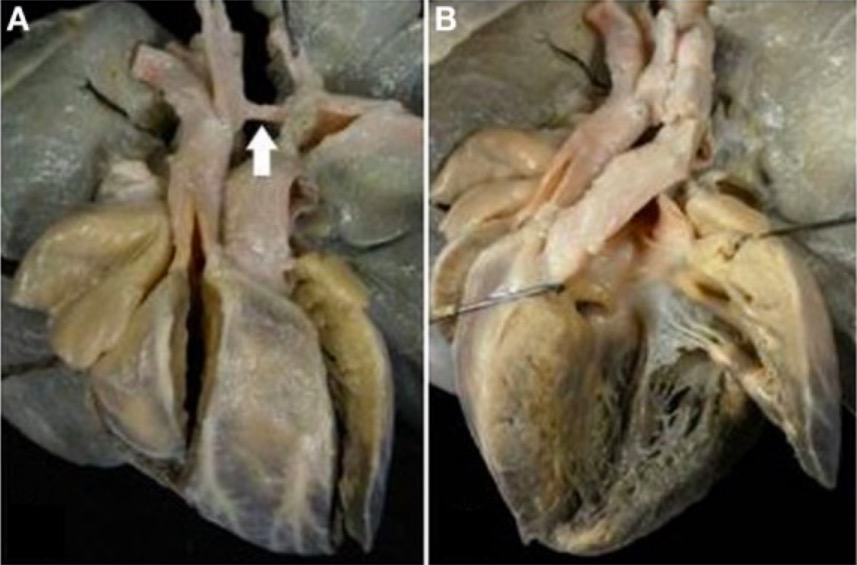
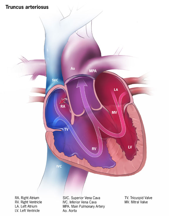
00:01 Let's review the five types of cyanotic heart disease. 00:05 We're going to go through each one. 00:07 We'll talk a little bit about each disease, so you can try and understand them a little better. 00:12 First, truncus arteriosus. 00:15 Truncus arteriosus - you can see here on your left side is a normal heart and on the right side is a patient with truncus arteriosus. 00:29 What you can see in this patient with truncus arteriosus is the aorta and the pulmonary artery have merged together and formed one main trunk, and that trunk is sitting over a VSD. 00:45 Here is the aorta and pulmonary artery with their common origin sitting over a VSD, and this VSD is allowing the blood from the left and right to mix together and then go up that common trunk. 01:03 In patients with this disease, they will have that common trunk with a single valve and it's supplying both the pulmonary and the systemic circulation. 01:15 You can see then why this baby would be born blue. 01:19 The truncus overrides the VSD, which results in that mixing lesion. 01:25 These patients are managed surgically, so we have to somehow repair this situation so that we've recreated a functional heart. 01:35 You can see the single trunk with a single valve is overriding both the pulmonary and the systemic circulation. 01:43 The VSD is allowing for blood mixture and is resulting in the baby being cyanotic. 01:50 Let's move on to transposition of the great arteries. 01:54 This is also called transposition of the great vessels. 01:57 In this condition, the aorta and the pulmonary artery are switched. 02:05 Blood in a normal patient will come back from the inferior and superior vena cava, down to the right atrium, to the right ventricle, and then out to the pulmonary artery, which goes to the lungs. 02:18 As you've learned, blood then comes back from the pulmonary vein, into the left atrium, the left ventricle, and then out the aorta to the body. 02:29 In a patient with transposition of the great vessels, these two main vessels have switched, which creates two parallel circulations. 02:38 In one of these patients, the blood comes from the inferior and superior vena cava, into the right atrium, the right ventricle, and it goes out to the aorta. 02:50 That blood now comes back from the body and goes back into the right atrium, the right ventricle, and into the aorta. 02:59 You can see how we don't ever get blood to the lungs that way, but they do because there's another parallel circulation going on where the blood is coming from the lungs, back to the left atrium, to the left ventricle, and out to the pulmonary artery. 03:19 This blood is circulating around to the lungs, and then you have another system that's circulating around to the body. 03:26 These patients will require a mixing lesion, such as a patent ductus arteriosus, or a VSD, or an ASD - and those mixing lesions will allow this patient to continue to survive. 03:44 That mixing allows some of the blue blood to get over the lungs and some of the pink blood to get over the body. 03:53 But you can see why in a hyperoxia test, this child would have a very, very low level of oxygen because boosting the amount of oxygenated blood in the pulmonary system is not going to necessarily improve the oxygen in the body. 04:07 You're really just relying on that mixing area. 04:10 In these patients, they need an arterial switch operation. 04:14 It's a bit complicated also and they usually require a catheterization because, remember, the coronary arteries can be coming off the left side or the aorta, or they can be coming off the right side, and you have to know where those coronary arteries are to fully fix the problem surgically. 04:34 Again, the right ventricle goes to the aorta, the left ventricle goes to the pulmonary artery, and the ductus, or the VSD, or ASD is keeping that patient alive. 04:48 Transposition of the great arteries is more common in infants of diabetic mothers. 04:57 This patient, if you get an x-ray, will have a classic egg on a string appearance where it looks like a sideways egg floating underneath a string with a very narrow mediastinum.
About the Lecture
The lecture Truncus Arteriosus and Transposition of the Great Vessels (TGV) by Brian Alverson, MD is from the course Pediatric Cardiology.
Included Quiz Questions
Which of the following features will allow a baby with transposition of the great vessels to remain alive at birth?
- Patent ductus arteriosus
- Mitral valve stenosis
- Pulmonary atresia
- Ventricular dilatation
- Surfactant administration at birth
Which of the following two structures merge together to form the defect of truncus arteriosus?
- Aorta and pulmonary artery
- Aorta and pulmonary veins
- Pulmonary artery and pulmonary veins
- Aorta and superior vena cava
- Pulmonary artery and superior vena cava
What is the first parameter of treatment in a case of truncus arteriosus?
- Surgical correction
- Observation
- Medication
- Angioplasty
- Heart transplant
Author of lecture Truncus Arteriosus and Transposition of the Great Vessels (TGV)

Brian Alverson, MD
Customer reviews
5,0 of 5 stars
| 5 Stars |
|
2 |
| 4 Stars |
|
0 |
| 3 Stars |
|
0 |
| 2 Stars |
|
0 |
| 1 Star |
|
0 |
Excellent lecture, good overview of the different CHD, thank you very much!
Great break down of each of the different cardiac conditions and how to differentiate between them!
