Playlist
Show Playlist
Hide Playlist
Formation of AV Canals

-
Slides 06-30 Formation of AV canals and Separation of the Ventricles.pdf
-
Download Lecture Overview
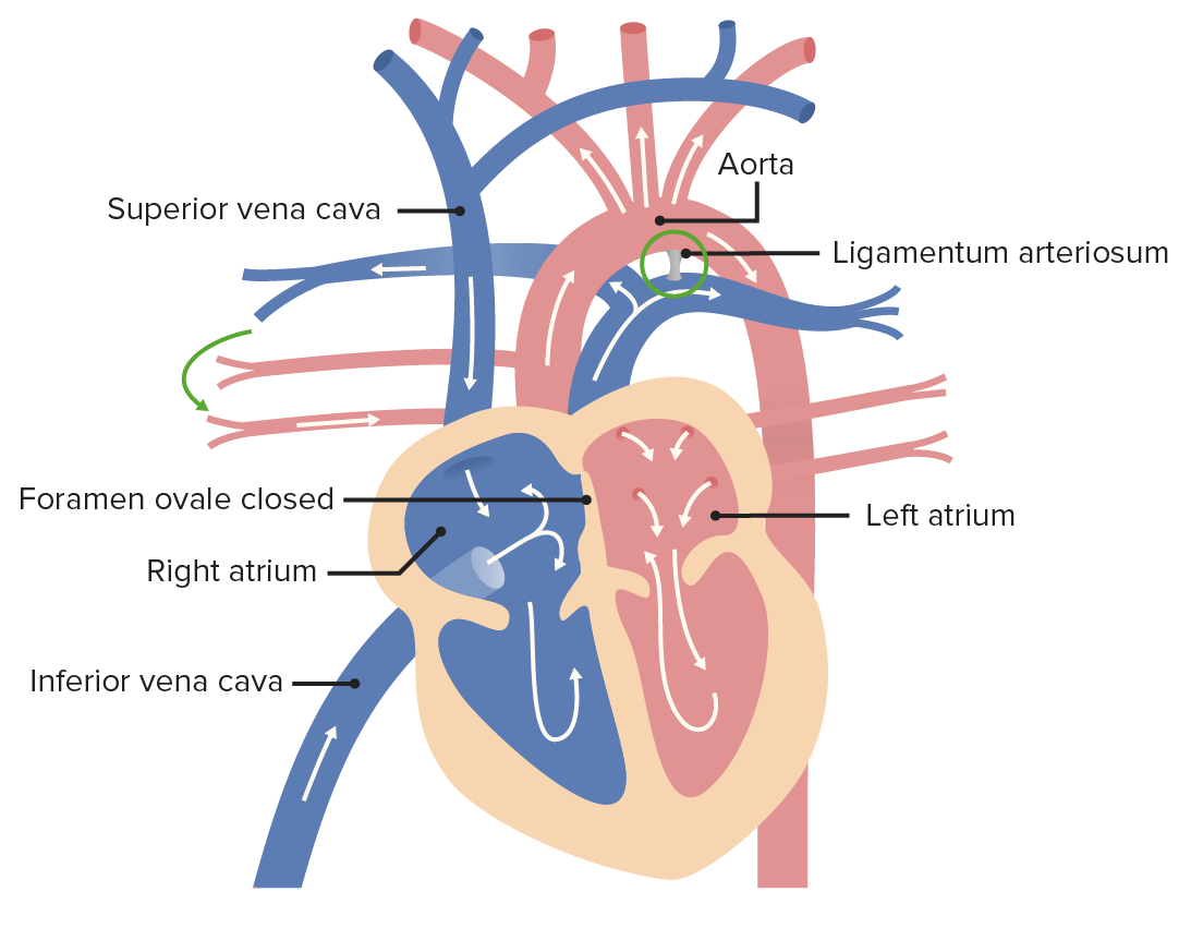
00:01 Hello and welcome back to our continuing discussion of formation of the heart. 00:05 Now we're gonna discuss how the atrioventricular canals and the ventricles separate to form the mature structures of the heart. 00:12 Just to put everything back into order, we're gonna start with the view of the heart here and we have the superior and inferior vena cava leading into the right atrium. 00:23 The right atrium is being separated from the left atrium by the septum primum, but they're still communicating with each other through the ostium primum. 00:31 Now thereafter, blood is gonna leave the atria and travel to the single ventricle which will then pump blood to the bulbous cordis and here we can see that we're starting to get a little bit of muscular upgrowth from the base of the ventricle towards the atrioventricular region and that's gonna be the first step in separating the two ventricles, right and left. 00:56 In the meantime, we have endocardial cushions on the right and left side where you got a right and left endocardial cushion and they're gonna be pinching together and narrowing the space through which the atria and ventricles are communicating. 01:11 And dorsally and ventrally' we can't see the ventral one because the front ventral half of the heart has been removed but we have a dorsal and ventral endocardial cushion that are growing closer together to occlude that midline portion of the atrioventricular canal. 01:27 So we go a little bit further and the ventral and dorsal cushions, also sometimes called the anterior and posterior endocardial cushions, have grown closer together and in the process they've narrowed the ostium primum that's connecting the left and right atria. 01:47 So each narrowing and the ostium secundum will be forming up in the septum primum of the atria, but as this narrowing occurs it's also closing the atrioventricular canals. 01:59 However, if it closed the atrioventricular canals completely we break our cardinal rule, we'd stop the flow of blood through the heart because blood could not leave the atria and travel to the ventricles. 02:11 So instead, we're gonna have to see how a single large open atrioventricular canal becomes two atrioventricular canals. 02:20 And this is actually fairly easy when you take a simplified schematic right here with the dorsal endocardial cushion on the top of the screen, ventral on the lower part and then right and left endocardial cushions indicated on either side. 02:33 The easiest thing to use as a way to conceptualize this is simply to make a circle with your fingers and then pinch it together to create two separate holes, and that's exactly what happens here. 02:47 The common atrioventricular canal gets subdivided into a right and left atrioventricular canal as the dorsal and ventral endocardial cushions grow together leaving two separate atrioventricular canals, one on the right and one on the left. 03:05 And the endocardial cushions form that continuous sheet of tissue that separates them as it closes the ostium primum in the atrium. 03:14 So if we take a view of the same process from the front ventral side of the heart, we can see with our view of the ventricle that the atrioventricular canal leaves the atrium and travels into the ventricle. 03:28 As development proceeds the cushions are starting to narrow the space of the atrioventricular canal. 03:35 Now once again, if it closed incompletely we'd interrupt the blood, so instead it leaves two separate openings where they're used to be one. 03:43 And we can see the blood is now able to leave the right atrium to get to the right side of the ventricle which will become the right ventricle, it can leave the left atrium and travel to the ventricle on the left side, and thereafter they're gonna leave through a common outflow tract. 03:59 At the same time, the muscular growth between the primitive ventricle and the bulbous cordis is growing towards the space with those endocardial cushions just closed. 04:11 So what can go wrong in this process? Well, essentially if your atrioventricular canal does not close completely, you have an atrioventricular canal defect. 04:21 A partial defect is going to mean incomplete closure of the right and left atrioventricular canals and this is usually associated with a ostium primum defect of the atrium because they didn't close, therefore, they didn't shut the ostium primum between the two atria. 04:39 It can also be associated with a ventricular septal defect on the lower side. 04:44 A complete atrioventricular canal defect however is a very serious problem because it means instead of a four chambered heart, you have a single chambered heart with venous blood entering the right atrium and just spilling into the left atrium, left ventricle and right ventricle and you're gonna have severe cyanosis and mixing of blood as this happens.
About the Lecture
The lecture Formation of AV Canals by Peter Ward, PhD is from the course Development of Thoracic Region and Vasculature.
Included Quiz Questions
Which of the following pairs of outgrowths will form the atrioventricular septum?
- Dorsal and ventral endocardial cushions
- Dorsal and left endocardial cushions
- Dorsal and right endocardial cushions
- Ventral and left endocardial cushions
- Left and right endocardial cushions
Which of the following describes a complete atrioventricular defect?
- Atrioventricular valves are not developed
- A low atrial septal defect with present atrioventricular canals
- A high ventricular septal defect with present atrioventricular canals
- Abnormal blood flow from the left atrium to the right atrium
- Abnormal blood flow from the right atrium to the left atrium
Author of lecture Formation of AV Canals

Peter Ward, PhD
Customer reviews
5,0 of 5 stars
| 5 Stars |
|
5 |
| 4 Stars |
|
0 |
| 3 Stars |
|
0 |
| 2 Stars |
|
0 |
| 1 Star |
|
0 |
