Playlist
Show Playlist
Hide Playlist
Lower Airways – Lung Anatomy


-
Slides 01 Respiratory Medicine Basics Brown.pdf
-
Download Lecture Overview
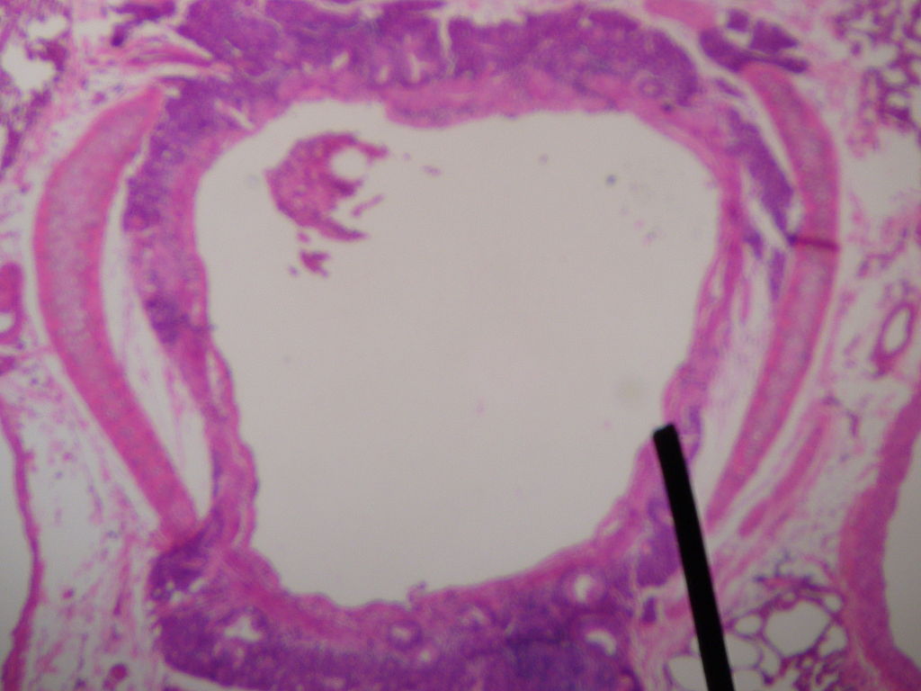
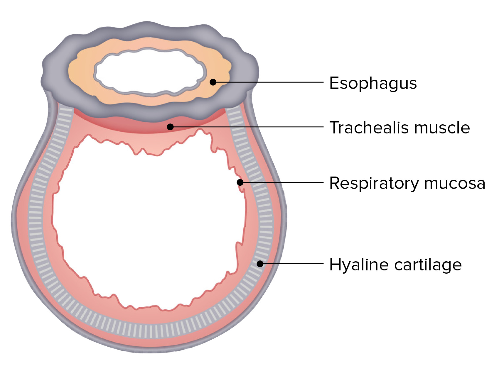
00:00 So that's the muscles and the skeletal system. 00:02 Now, we'll talk about the respiratory tract in more detail. We'll talk about the airways to start with. And the airways start, essentially, at the nose, but I'm going to talk about the lower airways only, which start at the larynx. And this starts with the trachea, which is a tube that runs from the larynx down to the division at the carinae into the right and the left main bronchi. And the order of the airways are: trachea, bronchi, these divide into bronchioles, terminal bronchioles, finally reaching the alveoli at the very bottom, at the very distal end of the lung. The airways are there to conduct air to the alveoli, and in the alveoli, the oxygen in the air gets into the blood, and the carbon dioxide in the blood gets into the alveoli. So the business end of the lungs is the alveoli, and the airways are all… are really there… just there to assure that air is supplied to the alveoli. 01:01 The trachea is a D-shape in cross-section, so posteriorly, there's a flat, tendinous bit. Anteriorly, there are cartilage curves with muscle and tendinous insertions... tendinous layers in between. It's about 11 cm long, a couple of centimeters in diameter. It run from the larynx right down to about the level of that 4th or 5th thoracic vertebra, where it divides into the right and to the left main bronchi, and that division is called the carina. The important thing about the trachea is that because it's running through the mediastinum—and the mediastinum is… are the structures between the right and the left lung—it's running next to blood vessels, thyroid gland, mediastinal lymph nodes, the esophagus, a variety of nerves, and that makes it at risk of any problems that are affecting those also affecting the trachea. So, for example, it's not uncommon for a very large mediastinal nodes to cause obstruction partially of the trachea. 02:02 After the trachea, you have the bronchi dividing into the right and the left lung. And the lungs themselves are very elastic, spongelike material consisting of very large numbers of alveoli. The right lung is slightly larger than the left lung. The contents of the lung are the bronchial tree, the alveoli, the blood vessels, the lymphatics draining the lung. 02:25 And importantly, they're surrounded by a thin layer of visceral pleura. All the vessels and all the air… the bronchi (the right and the left main bronchi) enter the lung through what we call are the hila, and these are gaps in the visceral pleurae which are very medial in the middle part of each lung. So the bronchial tree arises from the trachea. 02:52 It conducts air into each lung, and it also has incomplete cartilage rings just like the trachea does, although it's not a D shape now; they're more… they're round instead. 03:03 The bronchi branch successively to form smaller and smaller bronchi, and eventually, you end up with the alveoli. The main divisions, initially, of the bronchi are the right and left main bronchi. The left main bronchi then divides into a left upper bronchus and a left lower bronchus. The right side will divide into a right upper bronchus, and then there's an intermediate bronchus. And then the intermediate bronchus will divide into a right middle bronchus and a right lower-lobe bronchus. Each of those bronchi will feed air to a specific lobe. So on the right-hand side, there will be three main lobes: the right upper lobe, the right lower lobe, and the right middle lobe. And on the left-hand side, there would be a left upper lobe and a left lower lobe. The functional equivalent of the right middle lobe on the left side is called the lingula, and that is part of the left upper lobe. And these lobes are divided by invaginations of the visceral pleurae into the lung, and they form fissures—the main one being the oblique fissure, which is between the upper lobes and the lower lobe, with on the right-hand side being an additional fissure called the horizontal fissure, which divides the upper lobe from the middle lobe. 04:13 The bronchi, once they reach one lobe of the lung, will then divide, and there are segments for each lobe. And I'm not going to go into these in detail, because it's… we don't need to know precisely each segment. But here's a diagram showing each of the segments are present. And as you can see, they vary in number, depending on which lobe we're talking about. The smaller lobes, such as the right middle lobe, only having two segments, and the larger lobes, such as the lower lobe, having five or... four or five segments. And each of those segments has an individual bronchus feeding into it. And accompanying that bronchus, there's an individual pulmonary artery, pulmonary vein, and there'll be draining lymph nodes as well. The bronchi divide further and further in each segment, becoming tertiary bronchi and then small conducting vessels called terminal bronchioles, and these eventually become respiratory bronchioles. And one respiratory bronchiole will feed one gas exchange unit called an acinus. And that gas exchange unit will consist of alveoli, which is like small balloon-like objects opening off the terminal bronchiole and the alveolar ducts to form a cluster of air-filled sacs at the end of each bronchiole. 05:23 This is the business end of the lung. This is where gas exchange occurs. The important thing about the alveoli is that they are closely opposed to pulmonary capillaries, so each alveolus will have a lot of blood vessels—capillary blood vessels—surrounding it. And therefore, oxygen can move into the blood vessels from the alveoli relatively easily. So, for example, alveoli, about 7% of their surface is covered in pulmonary capillary. The numbers of alveoli are enormous. There's about 500 million in the average adult, and because of that, there's an incredible surface area of the lungs, allowing gas exchange to occur over a very large area. 06:03 And the average area is about 75 m2. Alveoli open off individual alveolar ducts, but there are the occasional connections between alveoli called the pores of Kohn. 06:17 If you look at the airway divisions—if you start at the trachea and then move down to the alveoli—there's about between 23 and 25 divisions before you get to an alveolus. 06:26 And the actual area of the lung increases exponentially as you go through those divisions, with the cent… with the trachea being 2 cm across, but then there's the 500 million alveoli with a surface area of 75 m2, and this is shown in this diagram. And you can see the exponential increase is mainly occurring in the lower airways. 06:44 This is a table giving the numbers of each individual air unit—air conductor unit—including alveoli, their rough diameter, and then, essentially, their cross-sectional area. And you can… and this basically gives you the data that the previous graph displayed, with the exponential increase in the surface area occurring when you get down to terminal respiratory bronchioles, the alveolar ducts, and the alveoli themselves.
About the Lecture
The lecture Lower Airways – Lung Anatomy by Jeremy Brown, PhD, MRCP(UK), MBBS is from the course Introduction to the Respiratory System.
Included Quiz Questions
Approximately how many airway divisions are there between the trachea and the terminal bronchioles?
- 25
- 15
- 20
- 30
In a transverse cross-sectional view, what is the shape of the trachea?
- D-shape
- C-shape
- Oblong
- Ovoid
- Circular
What is the approximate length of the trachea?
- 11 cm
- 5 cm
- 8 cm
- 9 cm
- 12 cm
At which vertebral level do the bronchi emerge from the trachea?
- 4th/5th thoracic vertebrae
- 2nd/3rd thoracic vertebrae
- 6th/7th thoracic vertebrae
- 8th/9th thoracic vertebrae
- 10th/11th thoracic vertebrae
Author of lecture Lower Airways – Lung Anatomy

Jeremy Brown, PhD, MRCP(UK), MBBS
Customer reviews
3,0 of 5 stars
| 5 Stars |
|
0 |
| 4 Stars |
|
0 |
| 3 Stars |
|
1 |
| 2 Stars |
|
0 |
| 1 Star |
|
0 |
Clear explanations, but more diagrams would be helpful in terms of visualisation
