Playlist
Show Playlist
Hide Playlist
The Neural Tube and Ventricular System


-
Slides 04-21 5 The Neural Tube and Ventricular System.pdf
-
Download Lecture Overview
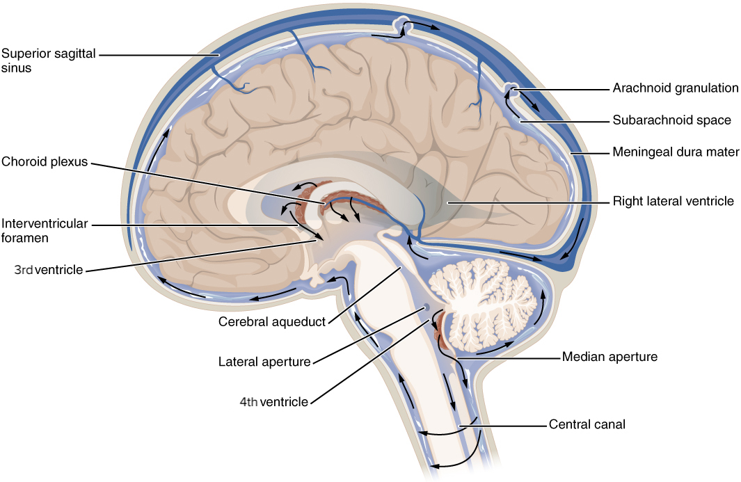
00:01 We´re now gonna turn our attention to the neural tube and more specifically the space within it. 00:07 That may seem relatively insignificant but the space inside the neural tube is vitally important to the normal function of the brain and the rest of central nervous system. 00:17 Now, initially there´s a neural canal at the core of the neural tube and at the cranial end where the brain and brain stem will be developing, it develops several bulges. 00:26 Initially, there are three primary vesicles that form: the prosencephalon which is going to be the forebrain, the mesencephalon, the midbrain and the rhombencephalon or the hindbrain. 00:40 So these three vesicles stage gives way to five vesicles. 00:45 The prosencephalon splits to become the telencephalon and the diencephalon. 00:51 The mesencephalon stays mesencephalon. 00:54 It´s still in the middle. 00:55 And then the rhombencephalon becomes the metencephalon and the myelencephalon. 01:00 Now, notice that the telencephalon has two bulges going off laterally that´s gonna be vitally important in just a moment because those are gonna form the lateral ventricles. 01:11 The telencephalon is the developing cerebral cortex and because we have a large left and right cerebral cortex, we have large lateral ventricles within them. 01:20 Within the diencephalon, which is going to be the developing thalamus, we´re going to have the third ventricle. 01:27 And the hypothalamus and thalamus are forming on either side of it. 01:32 The cerebral aqueduct is the portion of the neural canal that´s present within the mesencephalon. 01:39 The mesencephalon is gonna become the midbrain. 01:42 And then the fourth ventricle is the remnant of that neural canal that´s going to be present around the both metencephalon and the myelencephalon. 01:53 Now, the metencephalon will become the pons and the cerebellum and the myelencephalon is going to become the medulla oblongata. 02:01 Now, the rest of the neural canal stretches inferiorly along the developing neural tube and it will become the central canal of the spinal cord. 02:09 So this hollow space is going to be what forms our ventricular system. 02:15 So the ventricular system in the adult is based around the same structures. 02:20 The prosencephalon gave way to the telencephalon with its lateral ventricles and then the third ventricle, cerebral aqueduct, fourth ventricle and the rest of the central canal completely surrounded by the central nervous system. 02:35 The lining of these spaces is called the ependymal lining and it´s covered by ependymal cells that develop off of the neuroectoderm. 02:44 So the lateral ventricles are going to drain fluid into the third ventricle. 02:51 From there, the third ventricle fluid is gonna drain down the narrow cerebral aqueduct of the midbrain. 02:58 Then that CSF, cerebral spinal fluid, gets to the fourth ventricle which is surrounded by the cerebellum posteriorly and the pons and medulla anteriorly. 03:09 And lastly, the central canal of the spinal cord is presently extending all the way down to its end. 03:16 So what is it that creates the cerebrospinal fluid? The ependymal lining has small areas that start to enlarge and proliferate and create a structure called the choroid plexus. 03:28 Now, the choroid plexus is very vascular. 03:31 There´s a lot of blood vessels within it and it´s going to filter the blood and release the CSF into the ventricular system. 03:39 So the CSF is just an ultrafiltrate of blood and it´s gonna move through the ventricular system, but that´s a problem. 03:46 If it´s stuck in the ventricular system, it´s only gonna cause it to swell so it needs to have an exit and the exit is going to be through a portion of the medulla, pons and cerebellum where we have three little holes: the canals or the foramen of Magendie and Luschka and that allows the CSF to surround the brain and not just be found inside the ventricular system. 04:16 Now, let´s return to the outside for a moment and look at formation of the central nervous system overall. 04:25 Early on the tube is just a tube but at the three vesicle stage we developed a cervical flexure between the spinal cord and the rhombencephalon. 04:35 We also developed a cephalic flexure between the prosencephalon and the midbrain or the mesencephalon. 04:44 So those folds in the sagittal plane create a little bit of a kink in the developing neural tube. 04:51 As you move a little further in development we get there´s another one, the pontine flexure, that´s occurring between the myelencephalon and the metencephalon. 05:00 And this is one reason that our brains don´t go straight up and down from our spinal cord but have a little bit of a tilt as we are looking at it from a lateral or sagittal view. 05:10 Now, what can go wrong in this process? If we have interruption of ventricular system´s drainage we can develop a variety of conditions called hydrocephaly or hydrocephalus. 05:23 If we have blockage of the ventricular system it can cause swelling of the ventricles or pressure to be exerted on the brain. 05:31 Now, this occurs when we´re an adult. 05:33 The brain is surrounded by a very solid skull and that pressure has nowhere to go but to kind of press in and compress the brain and this is a medical emergency. 05:43 If we´re a neonate or an infant at that time, the brain swells but because the bones have not yet completely ossified it actually causes the head to swell as well. 05:55 So this hydrocephaly is a very notable thing if you see it in an infant. 06:01 So in adults, it´s a medical emergency that can cause very quick death and you may have to place a shunt to alleviate some of that pressure and if you note the pathologic specimen on the right, you can actually see the shunt has been placed inside the ventricles to drain that excess fluid. 06:18 They typically weave that down to the subcutaneous tissue into the peritoneal lining so that it can be drained into your body cavity and absorbed there. 06:26 Now, one thing to distinguish that from is cerebral aqueduct stenosis. 06:33 If your cerebral aqueduct inside the midbrain is too narrow, it can have a very difficult time getting fluid from the lateral ventricles and third ventricle to the fourth ventricle. 06:44 And remember, it´s in the fourth ventricle that the cerebral spinal fluid is able to escape the ventricular system and surround the brain and allow it to be cushioned so that every movement that I make doesn´t cause my brain to slam into my skull. 06:57 So if you have stenosis or narrowing of that canal, any swelling inside there can seal off your lateral and third ventricles and one problem with choroid plexus is it keeps producing cerebral spinal fluid all the time and if it keeps producing it and there´s no outlet, those ventricles, the lateral and third, will swell and put pressure on the brain from the inside and that is a medical emergency if it happens acutely. 07:23 If it happens gradually, it can be sometimes a little difficult to catch but will eventually cause neurologic symptoms and be diagnosable. 07:31 Congenital hydrocephalus and congenital aqueduct stenosis remain the most common indication for the placement of ventriculoperitoneal shunts. Up to 50 percents of V P shunts are placed for one of these two indications. 07:47 Alright, thank you very much and we´ll catch you on the next talk.
About the Lecture
The lecture The Neural Tube and Ventricular System by Peter Ward, PhD is from the course Development of the Nervous System, Head, and Neck. It contains the following chapters:
- The Neural Tube and Ventricular System
- Defects of the Neural Tube Development
Included Quiz Questions
What is another term for the midbrain?
- Mesencephalon
- Telencephalon
- Metencephalon
- Diencephalon
- Myelencephalon
The cerebral cortex arises from which secondary brain vesicle?
- Telencephalon
- Mesencephalon
- Diencephalon
- Metencephalon
- Myelencephalon
The pineal gland arises from which secondary brain vesicle?
- Diencephalon
- Mesencephalon
- Metencephalon
- Myelencephalon
- Telencephalon
What ventricular system disorder is characterized by an expansion of the lateral and 3rd ventricles, accompanied by a normal-sized to slightly collapsed 4th ventricle?
- Cerebral aqueduct stenosis
- Hydrocephalus
- Choroid plexus cyst
- Ventriculitis
- Normal-pressure hydrocephalus
Author of lecture The Neural Tube and Ventricular System

Peter Ward, PhD
Customer reviews
5,0 of 5 stars
| 5 Stars |
|
5 |
| 4 Stars |
|
0 |
| 3 Stars |
|
0 |
| 2 Stars |
|
0 |
| 1 Star |
|
0 |
