Playlist
Show Playlist
Hide Playlist
Limb Development and Muscle Migration

-
Slides 03-09 Limb Development and Muscle Migration.pdf
-
Download Lecture Overview
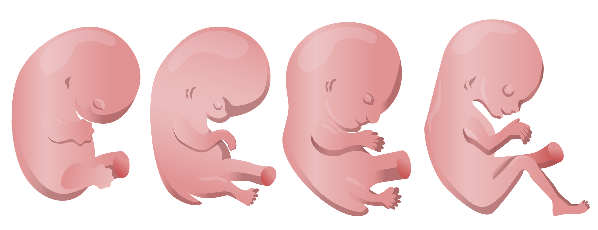
00:01 Hello. We´re gonna be discussing limb development and migration of the muscles as the limb buds form. 00:07 Now initially, the embryo has no limbs but on day 24, we have a small extension that´s gonna be the upper limb bud. 00:15 The lower limb bud follows a couple days later on day 26 and they´re gradually going to enlarge as development proceeds and give rise to the upper and lower limb. 00:24 Now, initially, there is a tail segment present between the two lower limbs but it initially gets displaced and then, the two lower limbs grow and the tail segment disappears. 00:34 As the upper and lower limbs extend, they´re gonna start to form a hand plate. 00:40 And that hand plate and the corresponding foot plate on the lower limb are going to have little rays of tissue that become more and more solid inside the little flipper-like hand plate that initially appears. 00:52 Those solid areas are called digital rays and the webbed tissue between those digital rays starts to thin and eventually, undergo apoptosis and die off and by the time we get to about day 52 or 56, those digital rays are very distinct and we have actual fingers and toes, 52 days for the upper limb and as usual, the lower limb lagging a few days, day 56, the toes are usually very distinct. 01:20 Now, as the limbs are extending, the upper and lower limb undergo a differential rotation that brings about the normal confirmation we have in the adult. 01:30 So as the lower limbs are growing and the upper limbs are growing, the upper limbs tend to rotate so that the elbow is pointing posteriorly. 01:38 That puts all of our flexor muscles anterior. 01:41 But the lower limb has the opposite happen. 01:43 The limbs rotate so that the knee winds up pointing anteriorly and all of our extensor muscles are on the anterior surface of the lower limb. 01:51 So that rotation difference between the upper and lower limb is what brings about that difference between extensors and flexors in the upper and lower limb. 02:00 So here we can see, they look relatively symmetric. 02:04 Then, the upper limb starts rotating and lagging a few days behind, the lower limb rotates with the elbow pointing posteriorly in the upper limb and the knee pointing anteriorly in the lower limb. 02:18 Now, as this is happening, the limb buds are not just full of loose tissue, they´re actually gonna be full of tissues that are becoming bone and muscle. 02:28 And the muscle overwhelmingly comes from the myotome which we recall comes from the somites on either side of the developing neural tube. 02:36 Now, the myotomes receive innervation from the developing spinal cord and as they migrate, they´re gonna form a few different structures. 02:43 One is gonna be the epimere. 02:46 The epimere is the cluster of developing muscle from the myotome that goes into the back and it´s going to become the intrinsic muscles of the back like the erector spinae. 02:55 The hypomere on the other hand is gonna travel into the torso, into the anterior thoracic and abdominal wall and also, into the limbs. 03:05 So the hypomere, pulling its nerve supply behind it is gonna migrate into the limbs and the body wall, the trunk. 03:14 Now, this is not happening in isolation as these are migrating, they pull their nerve supply behind them. 03:21 And so, the nerves are getting stretched away from the spinal cord as the upper and lower limbs are developing and those nerves get pulled behind making the brachial plexus in the upper limb and the lumbosacral plexus in the lower limb. 03:34 Now, this illustration is demonstrating that the muscles of the hypomere that migrate into the limb buds separate yet again and we have a posterior mass and an anterior mass or dorsal and ventral masses. 03:51 The ventral masses are gonna become the flexor muscles and the dorsal muscle mass will become the extensor muscles and they are gonna take up residence on either side of the developing bones of the limb. 04:04 Now, the nerves that are pulled along initially, are in a segmental layout. 04:09 You´ve got C4 to T2 contributing nerves to the myotomes that migrate into the upper limb and L2 to S3 contributing to the muscles that are gonna innervate and travel into the lower limb. 04:22 As the limb buds extend, those dermatomes and myotomes get stretched out separated by a ventral axial line. 04:30 So in this illustration you can see in the upper limb, C3, 4, 5, 6 and 7 are going to one direction, and then, they turn the corner and C8, T1, and T2 are taking care of the innervation on the inferior side of the upper limb. 04:45 A similar thing happens in the lower limb but as the limbs extend that easy kind of confirmation of the dermatomes and myotomes breaks down and we have the more complex dermatome map that we´re used in seeing in anatomy and clinical skills. 05:02 But one thing to note developmentally is that ventral axial line stays in place and on the anterior surface of the upper limb and on the posterior surface of the lower limb, that ventral axial line still separates those dermatomes. 05:16 Now, the complexity of those dermatomes is mirrored in the muscular nerve supply within and because these muscles are migrating, interdigitating, and weaving back out, that´s what´s gonna create the complexity of the brachial plexus and its various divisions and the lumbosacral plexus with its ventral and dorsal divisions going into the various muscle masses there. 05:43 Now, we´ve covered the muscles and the nerves but we mustn´t leave out the vasculature. 05:49 The vasculature for the developing limbs is coming from a segmental artery of the dorsal aorta. 05:54 Now, in the upper limb that will become the axillary artery and in the lower limb, the femoral artery. 06:01 Those are the two vessels that provide most of the blood to the limb. 06:04 Initially, the arteries go into the very deep substance of the limb bud and as it travels through, capillaries form and the venous drainage is more superficial. 06:15 So as these arteries extend, enlarge, they tend to stay very deep. 06:22 We don´t have very superficial arteries at the core of our limbs. 06:25 However, if you think about the cephalic, the cyclic veins in the upper limb, and the greater saphenous and small saphenous veins in the lower limb, you can see that there are still superficial veins and that´s a remnant of the fact that we had the venous system1 at a very superficial location early on during development. 06:45 So even though we have deep veins form alongside the arteries, those superficial veins do stick around as the arteries and veins develop. 06:53 Now, what can go wrong in this process? Sometimes, you can have failure of muscle migration or you can have flat out muscle agenesis where muscles don´t form at all. 07:04 This is relatively common in Poland Syndrome where you have an absence of one or more of the pectoral muscles. 07:10 That´s what´s shown in the picture on the left. 07:13 This child has an absence of the pectoral muscles on the right side. 07:17 And another fairly typical presentation of muscle agenesis comes in prune belly syndrome and it´s called that because there´s a failure of the abdominal musculature to form and these children tend to have not only severe muscular problems but also, renal problems but if they´re able to get a renal transplant, at that point, the loss of musculature in the anterior wall can be one of the major problems and they can actually do muscle, not transplants, but muscle flap procedures where they move a muscle from one place onto the abdominal wall and give these people a better quality of life. 07:52 So prune Belly Syndrome and Poland Syndrome both involve muscle agenesis but when you have a major developmental problem like that, it´s not likely that that´s the only problem. 08:03 It´s just a symptom of an underlying genetic defect that has problems manifesting in other places as well. 08:10 Sirenomelia is a disorder that is characterized by the presence of a single lower limb, formed of diffuse two lower limbs. 08:18 The most likely cause is unknown. 08:21 But the condition appears to be random, and it's considered very rare. 08:25 The condition is most likely related to impaired vascular development during the development of the lower limbs. 08:32 This could be triggered by genetic factors or environmental factors. 08:36 The most common environmental factor is maternal diabetes mellitus. 08:42 On the other extreme, occasionally, the tail segment can remain and people can be born with short non-functional tails. 08:50 Sadly, they´re not prehensile and terribly useful, they tend to be relatively floppy. 08:56 But if you have a vestigial tail that´s a remnant of the tail segment that should´ve rescinded but did not. 09:03 Last but not least, normally, we have the digital rays with the webbing in between undergoing apoptosis. 09:12 If that webbing incompletely goes away or doesn´t go away at all, you can have syndactyly or fusion of the digits. 09:22 On the right side of this upper picture we can see that the second and third digit have a bit of webbing between them. 09:28 This lower picture´s demonstrating a bit more significant fusion of the second and third digits of the lower limb and in this case, there´s likely some combination of the bones. 09:39 So syndactyly can involve a simple webbing between nearby digits but also, a fusion of the bones at the core of those digits and we can see here, there´s likely some degree of fusion of the proximal and maybe, middle phalanx of each one of those toes. 09:54 But the two distal phalanx are going to separate, giving us just tiny little nubs at the second and third toe. 10:03 Alright, thank you very much for your attention.
About the Lecture
The lecture Limb Development and Muscle Migration by Peter Ward, PhD is from the course Development of Musculoskeletal System and Skin.
Included Quiz Questions
What muscle group derives from the epimere?
- Intrinsic muscles of the back
- Muscles of upper extremities
- Muscles of lower extremities
- Muscles of the abdomen
- Facial muscles
Failure of muscle development during embryogenesis results in several sets of developmental anomalies. What muscle fails to form in Poland syndrome?
- Pectoralis muscle
- Abdominal muscle
- Biceps muscle
- Deltoid muscle
- Quadriceps muscle
Prune belly syndrome is a developmental anomaly that presents a failure to form which of the following?
- Abdominal muscles
- Pectoralis muscle
- Biceps muscle
- Deltoid muscle
- Quadriceps muscle
Which of the following is associated with maternal DM?
- Sirenomelia
- Anencephaly
- Syndactyly
- Vestigial tail
- Congenital hypothyroidism
What congenital defect results from the failure of apoptosis of interdigital tissues?
- Syndactyly
- Polydactyly
- Sirenomelia
- Amelia
- Mesomelia
Author of lecture Limb Development and Muscle Migration

Peter Ward, PhD
Customer reviews
5,0 of 5 stars
| 5 Stars |
|
1 |
| 4 Stars |
|
0 |
| 3 Stars |
|
0 |
| 2 Stars |
|
0 |
| 1 Star |
|
0 |
Dr. Ward is excellent! I thoroughly enjoy his lectures and feel well-versed in the subject after watching and listening to his explanations. Thank you!
