Playlist
Show Playlist
Hide Playlist
Infective Endocarditis (IE): Signs and Symptoms
-
Slides InfectiveEndocarditis InfectiousDiseases.pdf
-
Download Lecture Overview
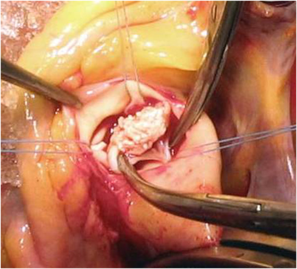
00:00 Well, how do patients present when they have endocarditis? You can expect fever in the vast majority. 00:07 A new murmur is heard in close to half. Now, let’s say you have been following your patient who’s had a heart murmur and you’ve known about it. Does that heart murmur change when they have endocarditis? The answer to that is usually not. Sometimes it does get worse, but usually not. So, it’s still going to sound mostly like the same old heart murmur. Some patients have microscopic or gross blood in their urine. Many patients with low-virulence organisms, it’s slow for them to go to the doctor and they may have fever for a couple of weeks. Well, this is where the spleen kicks in. 01:04 The spleen is the recipient of chronic bacteremia and may swell up in response to the bugs that are filtering through. So splenomegaly is common in endocarditis due to less virulent organisms. 01:24 More virulent organisms, on the other hand produce lesions like this. This would be referred to as a Janeway lesion. You’re going to find that it’s a flat, hemorrhagic lesion. This usually results from acute embolization of fragments from these vegetations. There are living organisms in these fragments. 01:48 So, these patients are often quite ill. In the old classification, these were the acute endocarditis types. 01:57 Also in the sicker patients, you may find something called Roth spots. Really what it is is as you look through the ophthalmoscope, you see retinal hemorrhages with a white center. So, it’s a white infarct of the retina surrounded by an areola of hemorrhage. There are other conditions in which you can find Roth spots. 02:25 You can find them in profound anemia. You can find them in leukemia. You can find them in certain connective tissue diseases like lupus. But when you see something like this, you certainly want to think of endocarditis and draw blood cultures. Unfortunately, the use of the ophthalmoscope is in decline for reasons that I’m not certain about. When you look at a typical work-up, you may see HEENT within normal limits, WNL. Now, I wonder if sometimes the WNL really means we never looked. 03:13 The emboli can show up also in the conjunctival sac. So, in somebody you’re working up for endocarditis, all you have to do is pull the eyelid down and you may see these conjunctival hemorrhages. 03:32 Osler’s nodes named for Sir William Osler, the actual founder of the specialty of internal medicine, wrote a treatise on infective endocarditis. Dr. Osler didn’t have any treatment that he could use for endocarditis but he described it better than most modern people could possibly describe it. 04:01 One of the things he found were these painful nodules that develop at the ends of the fingers and toes. 04:09 So, what’s happened is you had an emboli going to the fingers and toes from the vegetations usually due to lower-virulence organisms. The body then, the immune system surrounds these organisms and makes a little tender inflammatory nodule around them. That’s what you call an Osler’s node. 04:36 We’d like to say we find this in virtually every person but we see this in about 5% of patients. 04:49 Some other presentations that should get your antenna up are that of sepsis for no apparent reason, congestive heart failure for no apparent reason, a septic pulmonary embolus. This is common in a couple of situations. One, in IV drug users and if you think about it, they’re shooting things up via their arm veins. Of course, if those are infective things and contain staph in them then you will have organisms lodge in the lower lobes of the lungs and produce abscess. Also, those organisms gain access to the left side of the heart as well, or stroke in a young person. What’s that all about, especially if they have fever? You ought to think of endocarditis, or acute peripheral artery occlusion, or renal failure. I know of one patient that I saw when I was in my training. That was a young man. 06:01 He had a lot of acne but he came in with renal failure and he had no fever. So, there was nothing really to suspect endocarditis. But we found out that he had renal failure, had never had any predisposing conditions for it. So on a hunch, blood cultures were drawn and they were positive for coagulase, negative staphylococci, several blood cultures. So, patients in renal failure may not have the fever that you’re expecting. So, one of the causes of acute and chronic renal failure actually is infective endocarditis. As you might expect, these tiny little fragments from these vegetations go everywhere. That includes the brain. So, that leads to cerebral complications in 15% to 20% of patients. 07:11 They may have an ischemic or hemorrhagic stroke. But most of them have not had such a preceding diagnosis. They may have only a transient ischemic attack. It may be a totally silent embolism. Furthermore, you can have emboli that go to the vasa vasorum of the major blood vessels of the head. The vaso vasorum are the blood vessels that supply blood vessels. 07:47 If you embolized one of those, you would then weaken the wall of the blood vessel that supplied. 07:53 The infection can spread through the vessel wall and give something called a mycotic aneurysm. 08:01 That term mycotic is a misnomer. Mycotic seems to imply this is a fungal aneurysm. 08:08 Perhaps at one time, that’s what they were thought to be but actually these are most likely caused by bacteria. They also can embolize to the brain and result in a brain abscess or meningitis.
About the Lecture
The lecture Infective Endocarditis (IE): Signs and Symptoms by John Fisher, MD is from the course Cardiovascular Infections.
Included Quiz Questions
Which of the following lesions is associated with infective endocarditis and presents as painful, red, raised lesions found on the hands and feet?
- Osler's nodes
- Roth spots
- Janeway lesions
- Erythema nodosum
- Urticarial hemorrhages
What is the best definition of Janeway lesions?
- Small hemorrhagic lesions on the palms or soles caused by septic emboli from infective endocarditis
- Tense painful nodules at tips of fingers and toes from acute embolization of fragments of endocarditis vegetation
- Septic pulmonary emboli
- Ischemic stroke originating from infective endocarditis
- Hemorrhagic retinal infarcts caused by embolization from fragments of endocarditis vegetation
Which of the following signs is LEAST likely to be associated with infective endocarditis?
- Decreased deep tendon reflexes
- Murmur
- Congestive heart failure
- Hematuria
- Splenomegaly
Approximately what percentage of patients with infective endocarditis suffer from clinical cerebral complications, such as embolic strokes, mycotic aneurysms, or transient ischemic attacks?
- 15–20%
- 1–5%
- 80–90%
- 50–65%
- Less than 1%
Customer reviews
5,0 of 5 stars
| 5 Stars |
|
1 |
| 4 Stars |
|
0 |
| 3 Stars |
|
0 |
| 2 Stars |
|
0 |
| 1 Star |
|
0 |
I liked the clarity with which the topic was dealt with


