Playlist
Show Playlist
Hide Playlist
Pulmonary Clinical Anatomy: Introduction


-
Slides OverviewOnDyspnea RespiratoryPathology.pdf
-
Download Lecture Overview
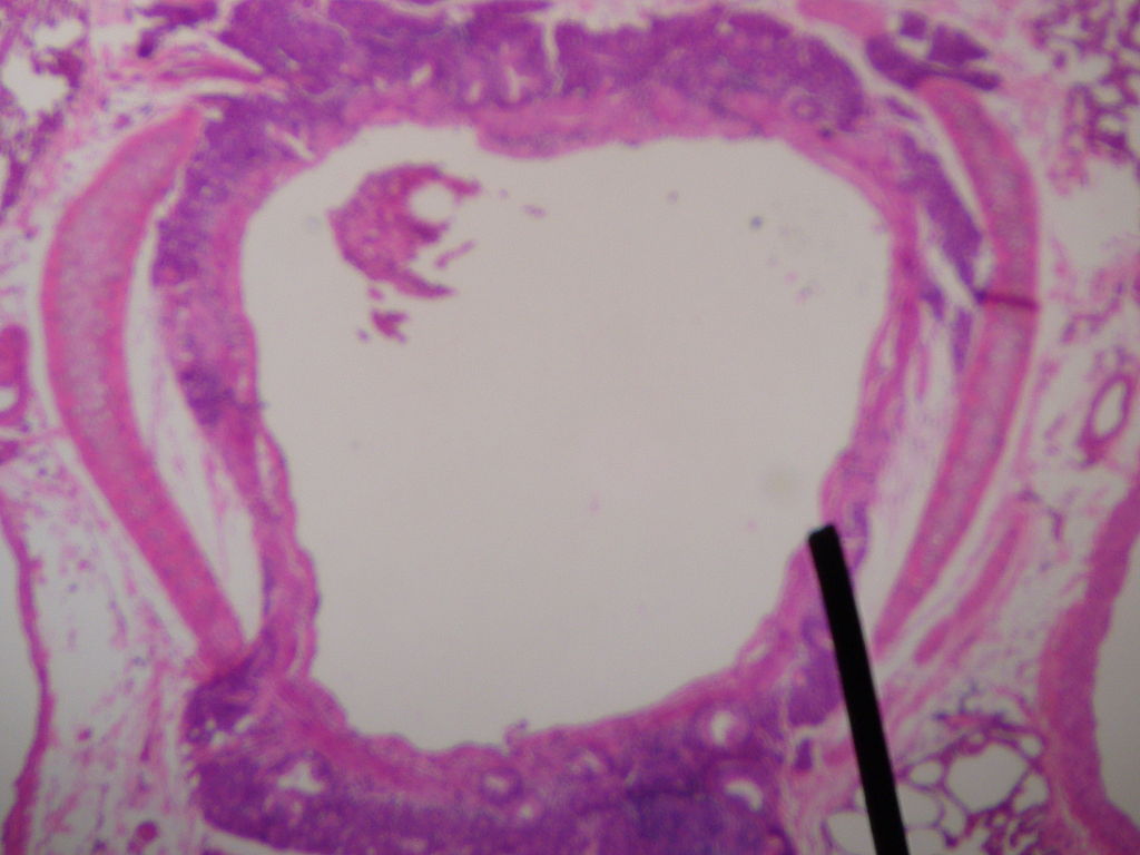
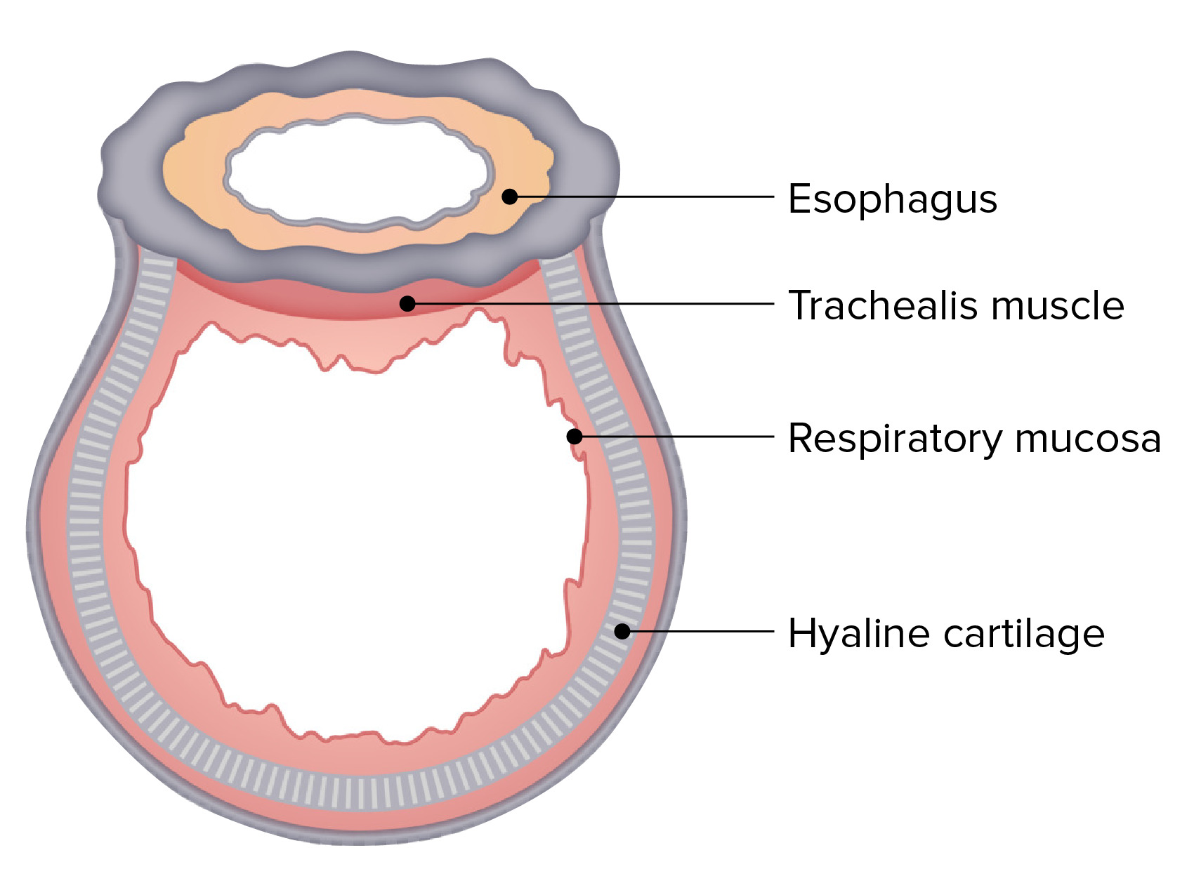
00:00 Welcome to Pulmonology. 00:03 Our approach in this lecture series will be the patient walked in through that door with signs and symptoms. 00:10 The patient walks in with cough. The history of that cough, "Was it a dry cough?" "Was it productive?" In terms of productive, what color was it? Was it stained as being brown? Was it rusty? Was it yellow? Was it green? Signs and symptoms is how you need to approach pulmonology here so that you have a broader view of what's going on with pathophysiology for each of the diseases and infections that we shall cover. 00:38 We'll walk through restrictive and obstructive diseases, but prior to any of that let's first take a look at signs and symptoms. 00:47 Overview of our bronchial tree. Begin at the proximal region with the trachea. 00:55 And then as we divide, divide, divide into branches or divisions, by the time you get all the way down to the alveoli distally, well you got to imagine as to how thin that alveoli is. 01:07 Right? And what about that trachea? The trachea is a supportive structure. It is basically an air tube. 01:15 That's exactly what it is. Meaning to say that it is then going to take the air that's coming in from the ambient air which has sea level is, what please. 01:25 Good, 760 mm as sea level. And you need to make sure that that trachea is nice and strong. 01:33 So therefore it is made up of cartilage or cartilaginous rings. 01:38 And then also, in the upper portion or the proximal portion of our respiratory tree, then we must have a method by which we defend ourselves against that ambient air. 01:49 Think about ambient air. There's a lot of stuff in there, it has antigens, it has allergens, so on and so forth. 01:56 So we need to make sure that we keep things like that out and so therefore think about the histology here as we branch deeper down into the alveoli. 02:04 That's important for you to understand and keep in mind. 02:07 I don't want you to just take a look at this and read what's on the Y axis or on the vertical or parallel words here. 02:15 That's just giving you an overview and things that you already know but what you're always bringing into play is "What is the function of the trachea? What kind of cells does it have?" It has mucociliary clearance. It has to be columnar cells. It has to be ciliated. 02:30 And the mucociliary clearance helps you take out any unwanted particles that you're breathing in, hhhmmm. 02:38 And you have to have mucous. Right? That's the proximal portion. 02:41 And then as you go further distally about the F cilia down in the alveoli, of course you don’t. 02:47 Why is the alveoli so thin? Type 1, type 2 pneumocytes are present. We know that it's squamous like, it has to be very thin. 02:57 Because what's across the alveoli membrane please? Exactly. 03:01 It's the pulmonary capillaries responsible for quite a bit of gas exchange. 03:05 So why would you want large columnar cells down there? Now, what does that mean to you pathologically? Now, what we shall do moving forward? Please understand. 03:13 And so we're going to plug in our infections into this respiratory tree. We're going to add in some diseases. 03:19 For example, we'll put in the most common lung cancer, adenocarcinoma. Isn't it? It is. 03:26 Adenocarcinoma is the most common. 03:27 But Dr. Raj, I thought that smoking was heavily, heavily associated with small cell lung cancer. That it is. 03:35 Or squamous cell cancer. That it is. 03:37 However, what if you're a non-smoker and could you still develop lung cancer? Sure, you could. 03:44 In the United States, it is the number 1 killer. 03:47 This is because lung cancer is the leading cause of mortality from cancer and the 6th leading cause of mortality overall according to the World Health Organization. 03:56 So therefore, we will have to know everything about bronchogenic adenocarcinoma. 04:00 So as we go through here, our doing here is setting up a nice little tree here and as we have in the proximal portion, these are cartilaginous and as you move further down into the bronchiole, the alveolar duct, and then in the alveoli. Hhhmmm. 04:16 The upper portion is known as a conducting zone. That's an important description that you need to know. 04:22 Remember all these from anatomy and physiology? Right? And the conducting zone literally is conducting air from the outside world down into the trachea. 04:30 And then as you move distally beyond the alveolar duct and you get into the respiratory zone. Makes perfect sense. 04:37 What's the respiratory zone responsible for? Gas exchange hence the name respiratory.
About the Lecture
The lecture Pulmonary Clinical Anatomy: Introduction by Carlo Raj, MD is from the course Introduction to Pulmonary Pathology.
Included Quiz Questions
What is the pressure of the atmosphere at sea level?
- 760 mmHg
- 250 mmHg
- 700 mmHg
- 540 mmHg
- 820 mmHg
Which of the following cells are responsible for the mucociliary clearance of the trachea?
- Ciliated columnar cells
- Non-keratinized squamous cells
- Cuboidal cells
- Pseudostratified cuboidal cells
- Ciliated pseudostratified squamous cells
Which of the following is NOT a part of the conducting system of the pulmonary tree?
- Alveoli
- Trachea
- Bronchi
- Bronchioles containing goblet cells
- Cartilaginous bronchi
Author of lecture Pulmonary Clinical Anatomy: Introduction

Carlo Raj, MD
Customer reviews
1,8 of 5 stars
| 5 Stars |
|
1 |
| 4 Stars |
|
0 |
| 3 Stars |
|
0 |
| 2 Stars |
|
0 |
| 1 Star |
|
4 |
It is a good introduction to anatomy and histology of respiratory tree.
4 customer reviews without text
4 user review without text
