Playlist
Show Playlist
Hide Playlist
Extravascular Hemolysis

-
Slides HemolyticAnemia RedBloodCellPathology.pdf
-
Download Lecture Overview
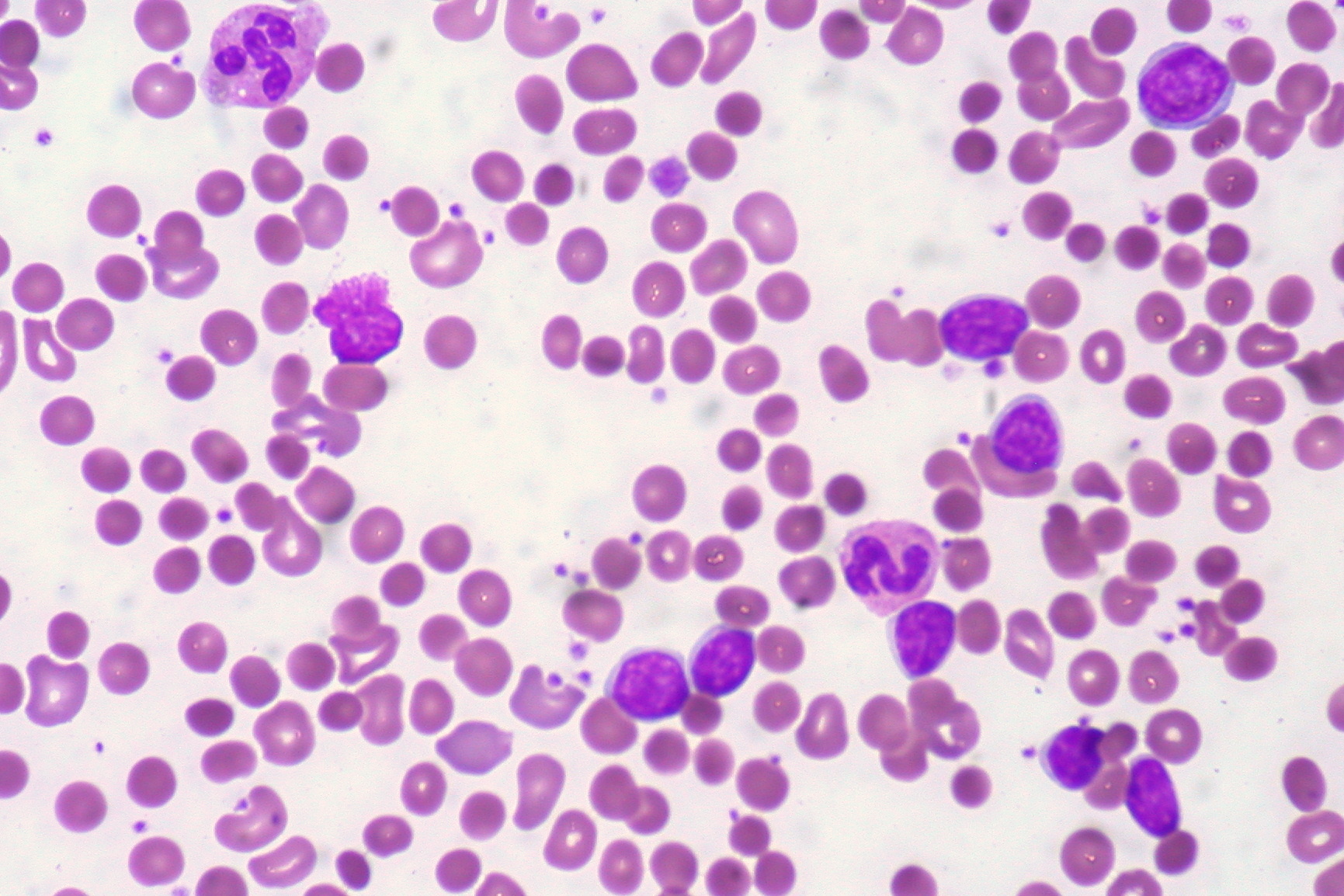
00:01 So, this is a nice little picture here to show you extravascular hemolysis. 00:04 Close your eyes, who’s your patient? Is it significant hemoglobinuria or is it significant jaundice? Good. 00:11 Significant jaundice. 00:13 Let me set this up. 00:16 We’re at the spleen, so therefore here’s my sinusoid of a normal RBC. 00:20 If it’s a normal RBC, what’s the lifespan of an RBC? 120 days. 00:26 And so therefore, if you have an RBC that has central pallor as you see here, well it’s not going to be destroyed by the spleen prematurely. 00:33 However, let us now go into the cords of Billroth, where are you? You’re in basically -- where the spleen and you see the guests. 00:44 What guests? You see these green cells that we see on the lining, these are the guests that you invited to dinner and they are hungry. 00:53 And so now, what are you going to serve them? The appetizers. 00:56 What are these appetizers? Oh, well, I’m going to serve you our famous dish known as sickle cell. 01:04 And so now, what happens? These RBCs are going to crazy because they love sickle cell. 01:09 No, you didn’t have to make this. 01:10 The point is you have a cell that becomes sickled. 01:15 It will be taken out of your vasculature, taken to the spleen. 01:18 So therefore, what kind of hemolysis? Extravascular. 01:22 Can you tell me as to what your sickle cell patient is going to look like? Good. 01:25 Jaundiced, icteric. 01:28 Now, jaundice can quite difficult to see in Africans, correct? And so therefore, you’re really looking for that icterus, is that clear? Next, well, oh my goodness, you really didn’t have to make this. 01:40 You made a spherocyte? It’s my favorite. 01:43 So now, you have a splenic macrophage which is going crazy on a spherocyte. 01:48 Once again, tell me about hereditary spherocyte. 01:51 Good. 01:51 Jaundice, icterus. 01:54 Are you seeing a pattern now? So you’ll notice, please, with your sickle and spherocyte, that is not what a normal RBC should look like. 02:02 And so therefore, these then will be destroyed by the spleen very, very quickly. 02:06 And the last one over here that you’ll see is an IgG, and later on, we’ll talk about what’s known as your autoimmune hemolytic anemia. 02:14 And what does an IgG mean? Well, this is desert? Really, you didn’t have to make this. 02:19 How did you know that I like fondue? What does that mean? IgG and C3b, you’ve learned in immunology that these are opsonins. 02:26 What’s an opsonin? It’s taking these “fruits,” maybe a strawberry, you dip it into chocolate. 02:35 That chocolate will be the opsonin. 02:36 What are the opsonins? IgG and C3b. 02:39 Well, some people might not like strawberries, but oh boy, you dip it into chocolate, it’s their favorite. 02:45 So now, you’ve made the RBC really tasty. 02:51 For whom? The splenic macrophage. 02:52 Guess what kind of hemolysis? Good. 02:56 Extravascular. 02:56 Are you seeing the pattern here? Understand where we are and what the disease is. 03:02 Ultimately, understand who your patient is. 03:04 All jokes aside, this is a serious matter. 03:06 So the bottom portion here is that you’re going to be releasing unconjugated bilirubin. 03:13 What is the laboratory term that we use in medicine? U-N is I-N. 03:17 Indirect. 03:18 Which means what? Lipid soluble. 03:22 And you would expect to find lactate dehydrogenase, but that is going to be nonspecific. 03:25 You’ll find that both in intra and extravascular. 03:29 Make sure you spend a little bit of time and you understand this in its completeness. 03:31 Now, let’s move on.
About the Lecture
The lecture Extravascular Hemolysis by Carlo Raj, MD is from the course Hemolytic Anemia – Red Blood Cell Pathology (RBC).
Included Quiz Questions
What is the approximate lifespan of normal red blood cells?
- 120 days
- 60 days
- 180 days
- 160 days
- 80 days
Which of the following is the main product of extravascular hemolysis?
- Unconjugated bilirubin
- Conjugated bilirubin
- Direct bilirubin
- Biliverdin
- Albumin
Which of the following does NOT indicate hemolytic anemia?
- Cholesterol gallstones
- Unconjugated bilirubinemia
- Low serum haptoglobin
- Hemoglobinuria
- Elevated lactate dehydrogenase
Author of lecture Extravascular Hemolysis

Carlo Raj, MD
Customer reviews
5,0 of 5 stars
| 5 Stars |
|
6 |
| 4 Stars |
|
0 |
| 3 Stars |
|
0 |
| 2 Stars |
|
0 |
| 1 Star |
|
0 |
Dr. Raj is so funny! How did you know that I like vondoo? I will never forget extravascular hemolysis lol. Thank you for a great lecture!
This was a funny video. I enjoyed it. Thanks for making the subject easy to digest :-)
Dr Raj is one of the best lecturers in Lecturio, I really like the way he explains everything and his sense of humour
Considerate and well performed explanations, look forward for more good outcomes. Thank you doctor.
