Playlist
Show Playlist
Hide Playlist
Mammalian Cell: Structural Components and Their Functions

-
Basic Histology 02.pdf
-
Download Lecture Overview
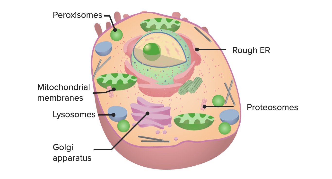
00:00 In this lecture, I'm going to describe structural components of the cell and their functions. 00:09 My name is Geoff Meyer. 00:12 I'm only going to cover the basic structures within the cell and their functions because I don't want this to be a full cell biology lecture or a molecular biology lecture. 00:25 It merely is to provide you with sufficient background information to then understand the histology of basic tissues and also organ systems that follow. 00:42 Here is a diagrammatic representation of a typical mammalian cell. 00:49 All the major structures that I will talk about in this lecture are labeled in this diagram. 00:58 You can see how complex the structure of a cell really is and all those different components you see labeled there have their own specific functions, although they work together to perform the various roles in which cells play in our various tissues and organs. 01:20 I'm not going to talk about the microvillus projections from the cell you see at the top of this image. 01:26 Those microvillus projections would be more important when we look at epithelial cells in later lectures. 01:34 But I am going to look at all the remaining structures you see labeled there. 01:42 First of all, let's look at the cell membrane shown in this image. 01:48 It looks a very complicated image, but focus firstly on the little round circular structures you see making up the bulk of this cell membrane. 02:02 It's a bi-layered membrane, and those little yellow circular structures you see are the phospholipid heads that are polar and hydrophilic so they spontaneously orientate towards the aqueous surface of the cell. 02:24 Whereas the fatty acid tails or chains you see, they're nonpolar, they're hydrophobic and therefore, they passively rotate towards the center of the bi-layer, away from the aqueous medium. 02:43 So that creates what you see there is this bi-layer of the cell membrane. 02:50 You can also see these rather colorful large structures also attached to the cell membrane. 03:00 These large structures represent proteins. 03:05 They can be transmembrane proteins such as the one you see down in the bottom left hand side that project right through the whole width of the cell membrane, which is about 8 nm to 10 nm in width, or you can have some integrated proteins you see that are attached to either the internal or the external surface of this bi-layer. 03:31 These proteins perform very important functions. 03:37 Some of them act to hold cells together to form intercellular junctions, and we'll see those when we look at epithelia in more detail in other lectures in this histology series. 03:53 Some of the proteins also act to create pumps or channels to allow ions and molecules to go in and out of the cell. 04:04 Other proteins serve as receptors. 04:07 Chemical messengers can attach to these receptors and initiate messaging within the cell and therefore excite function of the cell or inhibit functions of the cell. 04:21 And yet other molecules, other proteins you see, can act as enzymes on the surface of the cell and we'll see these in more detail when we look at the intestinal cells in lectures on the Intestinal System. 04:39 Other proteins can also act in cell recognition. 04:46 Also, what you see attached to some of these proteins are what we call the cell coat or glycocalyx. 04:55 These are glycoprotein complexes or glycolipids. 05:00 They're also involved in cell recognition, but they also help to separate one cell from its neighbor. 05:12 Now, here's a section through a piece of tissue. 05:17 It happens to be in the kidney of the human, and what I want to do is just initiate our talk of what's in a cell by just focusing on the structure that's running down from the top right hand side of the image to the bottom left hand side. The structure that has all these circular bluish stained structures. 05:45 They're the nuclei of cells. 05:48 And there's other cells you see in the periphery of the image that are also nuclei, but focus on the condensed numbers of nuclei in this central structure running down the image. 06:01 It happens to be a section through the wall of a tubule in the kidney. 06:08 Well, those circular structures as I've mentioned a moment ago is the cell nucleus and the clumpy stained material you see is chromatin. 06:21 I'm going to describe chromatin in a moment. 06:27 The pinker-stained region between these nuclei is going to represent the cell cytoplasm and it's within that cytoplasm that I'm going to describe all the important structural components. 06:44 The cell cytoplasm is bound on the external surface by a cell boundary and if you look very, very carefully in the middle of this section, in the middle of this tubular structure that I've described to you, you can just see a fine pink line running between the cells. 07:04 That's going to represent the cell boundary between one cell and its neighbor. 07:10 That's where those cell membranes or the plasma membrane is going to be. 07:17 And you can tell when you look at the cell boundaries roughly what the shape of these cells are. 07:23 They tend to be a more cuboidal shape rather than a columnar shape. 07:30 And another way in which you can tell that is to look at the nuclei. 07:34 Generally, when you see a nice round nucleus, it means that the cell is cuboidal. 07:40 If the nucleus was elongated, it would mean that the cell was columnar. 07:48 And then between the cells, you can see these white clear spaces, that's the interstitial space. 07:57 It's full of interstitial fluid that bathes the cells. 08:01 That's where the cell gets its nutrients from and exchanges products to and from the cell membrane into the cell itself. it's a bit exaggerated in this section because normally, in normal paraffin H and E stain slides or sections of tissue, you get a little bit of shrinkage of tissue and so you get some exaggeration of some of these spaces you see in cells. 08:30 We call that artifact. 08:33 It's a product of the processing. It's not a real product of the real structure of the tissue. 08:41 If you look carefully on the left hand side of the image, you see some bright-stained red blood cells. 08:49 Red blood cells are about 10 microns in diameter, so those little red blood cells can be a good ruler or a good guide to enable you to tell the size of some of the nuclei and the size of some of the cells you see in this particular section.
About the Lecture
The lecture Mammalian Cell: Structural Components and Their Functions by Geoffrey Meyer, PhD is from the course The Mammalian Cell.
Included Quiz Questions
Which of the following best describes the hydrophilic region of a cell membrane?
- Water-loving
- Water-fearing
- Nonpolar
- Lipid-soluble
- Hydrophobic
Which of the following forms the channels and pumps in the phospholipid bilayer?
- Proteins
- Lipids
- Carbohydrates
- Hydrophilic heads
- Hydrophobic heads
Which of the following is NOT a component of a cellular membrane?
- Nucleic acids
- Proteins
- Carbohydrates
- Phospholipids
- Glycoproteins
What makes up the tail of a phospholipid?
- Fatty acids
- Proteins
- Glycoproteins
- Nucleic acids
- Carbohydrates
How many layers are in a cell membrane?
- 2
- 1
- 3
- 4
- 5
Author of lecture Mammalian Cell: Structural Components and Their Functions

Geoffrey Meyer, PhD
Customer reviews
5,0 of 5 stars
| 5 Stars |
|
1 |
| 4 Stars |
|
0 |
| 3 Stars |
|
0 |
| 2 Stars |
|
0 |
| 1 Star |
|
0 |
excellent presentation explaining step by step important concepts at the right pace
