Playlist
Show Playlist
Hide Playlist
E-FAST Exam: RUQ, LUQ, Bladder & Pericardial View


-
Emergency Medicine Abdominal Injuries and Hemorrhagic Shock.pdf
-
Download Lecture Overview
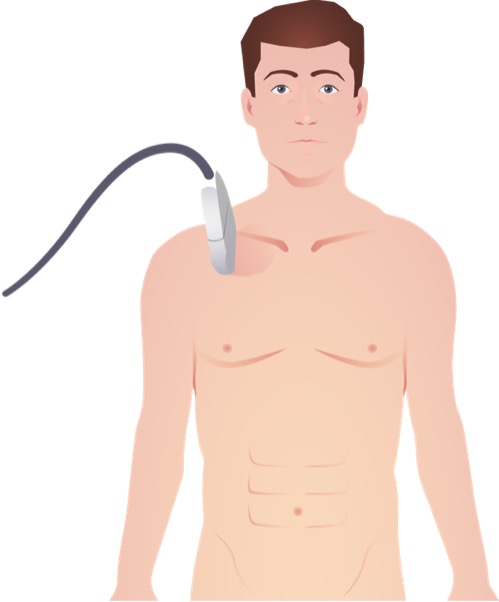
00:01 Alright, we already talked about the lung views of the FAST as part of the pulmonary trauma lecture, so we’re not gonna cover that here. 00:08 Now, I’m gonna talk about the abdominal and the pericardial views. 00:12 So here are some images of the right upper quadrant view. 00:15 We’re placing the probe at the patient’s subcostal area in roughly the anterior to mid axillary line and we are looking for the interface between the liver and the kidney. 00:30 This interface is known as Morison’s pouch and you can see the liver and the kidney are labeled on this image here. 00:37 If there is hemoperitoneum, you’re gonna see a black line separating the liver from the kidney. 00:44 Now in a normal, healthy person Morison’s pouch is a potential space, there’s nothing in it. 00:49 But if the patient has blood in their belly, then there’s gonna be blood that sort of intercalates down into Morison’s pouch and separates the liver from the kidney with a black stripe and you can see that labeled on this image. 01:02 This is abnormal and diagnostic of hemoperitoneum. 01:06 The left upper quadrant view is very similar to the right upper quadrant view except it’s done on the left hand side of the body. 01:13 The spleen is a smaller organ than the liver but otherwise looks similar on ultrasound. 01:19 And in this case, rather than seeing blood collecting in between the spleen and the kidney which you might see in the splenorenal recess in some cases, now we’re seeing blood sort of around the spleen underneath of the diaphragm. 01:31 Again, you can see where the blood is labeled on the image so instead of seeing the spleen right up against the diaphragm, you’re seeing it sort of floating in a black pool and that again is suggestive of hemoperitoneum. 01:43 Lastly, we have the bladder view where the probe is placed in the suprapubic region just over the bladder. 01:50 You can see the fluid-filled bladder at the top of the image there and then the area of blood underneath of it which is labeled on the image is abnormal. 02:01 Normally, the rectovesical space should not have any fluid in it and in this case we’re seeing a black stripe outlining the bladder which is suggestive once again of hemoperitoneum. 02:12 So these are the three views that give us information about whether there’s bleeding in the abdomen or pelvis. 02:17 The fourth view that we wanna think about is the pericardial view. 02:21 So this is a picture of the pericardial view. 02:25 What we’re doing is placing the probe under the xiphoid process and we’re pointing it up towards the left shoulder. 02:32 You can see the liver at the top of the image, the left lobe of the liver is it sort of extends past the midline in most patients and is actually used as a window to view the heart and we’re looking at the heart from the apex subs so you can see the ventricles first followed by the atria a little bit deeper. 02:49 And the really striking thing on this image is, again, labeled clearly here and that’s blood. 02:54 So there is a circumferential black stripe that outlines the heart. 02:59 This is not normal. 03:00 Normally, the pericardium is gonna be right up against the heart and there’s not gonna be any fluid in between the pericardium and the heart so you’re not gonna see this sort of a stripe, but when you see it that is diagnostic of hemopericardium in the trauma setting. 03:17 So what are we gonna do about it? Well, I mentioned that there’s a specific treatment that we need to perform for patients with hemopericardium and tamponade and that is emergency pericardiocentesis. 03:28 So the way we perform this procedure is by approaching the patient the same way that we did the ultrasound, we’re gonna go underneath of the xiphoid process with an 18 gauge spinal needle. 03:38 You’ll need a pretty long needle for this. 03:40 We’re gonna point it towards the left shoulder and aspirate continuously and you can use ultrasound guidance to make sure that you’re on the right location or if that’s not available to you in your setting you can also use ECG guidance when the needle tip is beginning to touch the epicardium, you’ll see ST segment elevations as depicted there on the top of the image. 04:02 So either one will tell you when you’re on the right general location. 04:05 And basically, you’re gonna be aspirating continuously until you get into the pericardial fluid collection and you’re gonna evacuate it. 04:15 If your patient has ongoing pericardial bleeding and you think you need to place a catheter for repeated drainage of the pericardium, you can actually use a guidewire in order to put a catheter in although typically in the immediate trauma setting, we’re gonna do this as a onetime procedure to stabilize the patient prior to sending them to the operating room for more definitive management. 04:37 Alright, so our take home points about hemorrhagic and other forms of shock and trauma are one, that this is a diagnosis that you make by monitoring the patient’s vital signs. 04:48 Vital signs correlate very well with the degree of blood loss in most, but not all cases. 04:54 Physiologic compensation can mask blood loss so for patients who are well compensated, you might not see signs and symptoms of blood loss until later so you need to be really vigilant and take even subtle vital signs abnormalities seriously. 05:09 We’re gonna always treat hemorrhagic shock with volume. 05:14 We’re never gonna use pressors to treat bleeding, that’s very important. 05:18 We wanna have adequate IV access. 05:20 We’re gonna start off with isotonic crystalloid and follow that with blood products as needed. 05:25 And we always wanna make sure that we identify the source of bleeding in patients with hemorrhagic shock as well as ruling out other injuries like hemopericardium or tension pneumothorax which can be accomplished with an E-FAST. 05:40 Thank you very much.
About the Lecture
The lecture E-FAST Exam: RUQ, LUQ, Bladder & Pericardial View by Julianna Jung, MD, FACEP is from the course Trauma (Emergency Medicine).
Included Quiz Questions
The Morison's pouch is a potential space where fluid can accumulate in abnormal conditions. Where is this potential space located?
- Between liver and right kidney
- Between spleen and right kidney
- Between liver and gallbladder
- Between pancreas and liver
- Between adrenal gland and kidney
What is the correct treatment for shock in a patient assessed to have an accumulation of blood around the heart during an eFAST scan?
- Pericardiocentesis
- Chest tube thoracostomy
- Thoracotomy
- Volume replacement
- Paracentesis
Which of the following statements regarding pericardiocentesis is TRUE?
- The goal of the procedure is aspiration of blood from the pericardium to permit cardiac filling.
- The procedure should be performed with a 16-18 gauge intravenous catheter.
- The needle should be inserted under the xiphoid process and aimed toward the right shoulder.
- CT scan should be used to confirm the diagnosis of hemopericardium before attempting the procedure.
Author of lecture E-FAST Exam: RUQ, LUQ, Bladder & Pericardial View

Julianna Jung, MD, FACEP
Customer reviews
5,0 of 5 stars
| 5 Stars |
|
5 |
| 4 Stars |
|
0 |
| 3 Stars |
|
0 |
| 2 Stars |
|
0 |
| 1 Star |
|
0 |
