Playlist
Show Playlist
Hide Playlist
Anatomy of the Ear esp. Ossicles

-
Slides 9 AuditorSystem BrainAndNervousSystem.pdf
-
Download Lecture Overview
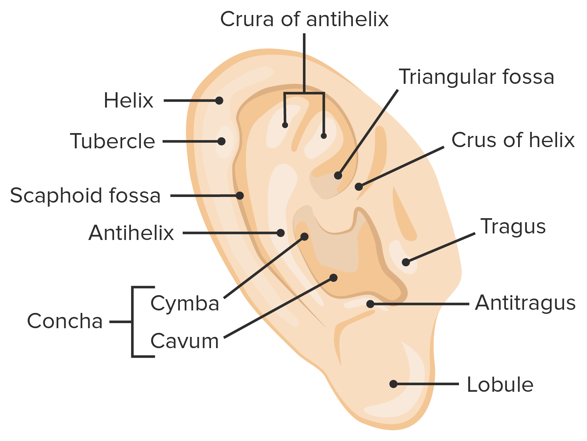
00:00 Welcome to this presentation on the auditory system. First, I want you to understand the basic components of the ear. We’ll begin with the outer ear. Everything shaded is a part of the outer ear, so the most prominent structure is external. That is the auricle. Then it will collect sound waves and then funnel those sound waves into the external acoustic meatus. Then those sound waves will strike the tympanic membrane or eardrum. The middle ear is shown in through here. It is characterized by the presence of the three very tiny ear ossicles. The middle ear will connect to the pharynx through the pharyngotympanic tube that we see here. Then lastly, we have the inner ear. The inner ear is going to house the machinery or the cellular architecture that’s responsible for the generation of action potentials that will allow us to perceive what we hear. There is a clinical application for you to understand with respect to the tympanic membrane. This procedure that we’re going to briefly describe in why you might want to do it is referred to as a tympanostomy. This is essentially the insertion of ear tube. If we take a look down here, you see the tympanic membrane. Then you have the tube or grommet right in through here allowing a communication then between the middle ear and the external ear. This procedure can be done if an individual, particularly a youngster has recurrent ear infections. This would then help reduce the pressure because of the fluid build-up in the middle ear. It would also allow the drainage of that fluid and any accompanying pus as a result of the chronic ear infections. Since the goal would be to eliminate the infection, this allows for antibiotic drops to be inserted into the ear canal, the external acoustic meatus, and then actually pass through the ear tube into the middle ear where they can be effective against the microbes that are causing the infection itself. The ear ossicles that are prominent features of the middle ear can be remembered by the can’t mis, M-I-S, mnemonic. This describes the order of the ear ossicles from the tympanic membrane to the oval window which is the entry or the transmission of sound waves into the cochlear apparatus. The M stands for malleus. We see the malleus highlighted here. 03:30 It does have a component that is attached to the tympanic membrane. So, when the tympanic membrane receives the sound waves, it will start movement of the malleus in the beginning of the ear ossicle chain. 03:47 The I in mis is the incus. Now we see it shaded. Then the final structure is the stapes that resembles a stirrup that we see shaded as well. The purpose of these ear ossicles is for the osseous conduction of sound waves. When those sound waves strike, the tympanic membrane, it starts to vibrate and then that causes the ear ossicles to vibrate as well. That vibration is conducted to the oval window and then into the cochlear apparatus. The ear ossicles have muscles associated with them that attach to them. 04:37 One such muscle is the tensor tympani. We see the sensor tympani here shaded. It will attach to the malleus. 04:48 It is innervated by the mandibular nerve which is V3. This is one of the nerves or divisions of cranial nerve number five, the trigeminal nerve. The stapedius is another muscle within the middle ear. 05:09 It attaches to the stapes. It is not shown in this illustration. It is innervated by a small branch of the facial nerve. 05:19 Though these muscles are very small in size, they do have a function in that when they contract, they will reduce the oscillations that occur between the ear ossicles. By doing that, they will attenuate sound wave conduction through the middle ear. If one or more are paralyzed, then one has an increased sensitivity to sound. That would be termed hyperacusis.
About the Lecture
The lecture Anatomy of the Ear esp. Ossicles by Craig Canby, PhD is from the course Auditory System and Vestibular System. It contains the following chapters:
- Ear – Components
- Ear Ossicles
Included Quiz Questions
What parts of the ear communicate with the eustachian tube?
- Middle ear and pharynx
- Middle ear and nasal cavity
- Inner ear and oral cavity
- Middle ear and frontal sinus
- Inner ear and pharynx
Which of the following statements regarding tympanostomy is FALSE?
- It is helpful in the removal of foreign bodies.
- It is indicated in recurrent middle ear infections.
- It can be used to administer antibiotics in the middle ear.
- It decreases the pressure in the middle ear.
- It can be used to drain pus and fluids.
Which of the following statements regarding ear ossicles is most accurate?
- The malleus has a component attached to the tympanic membrane.
- The incus is attached to the oval window.
- The tensor tympani muscle is attached to the stapes.
- The stapes receives signals directly from the tympanic membrane.
- The stapes communicates with both the malleus and the incus.
Author of lecture Anatomy of the Ear esp. Ossicles

Craig Canby, PhD
Customer reviews
5,0 of 5 stars
| 5 Stars |
|
1 |
| 4 Stars |
|
0 |
| 3 Stars |
|
0 |
| 2 Stars |
|
0 |
| 1 Star |
|
0 |
I recommend all the lectures to all IMGs out there to brush up your knowledge on your basic subjects.
