Playlist
Show Playlist
Hide Playlist
Bony Anatomy in Radiology
-
Slides Projections.pdf
-
Download Lecture Overview
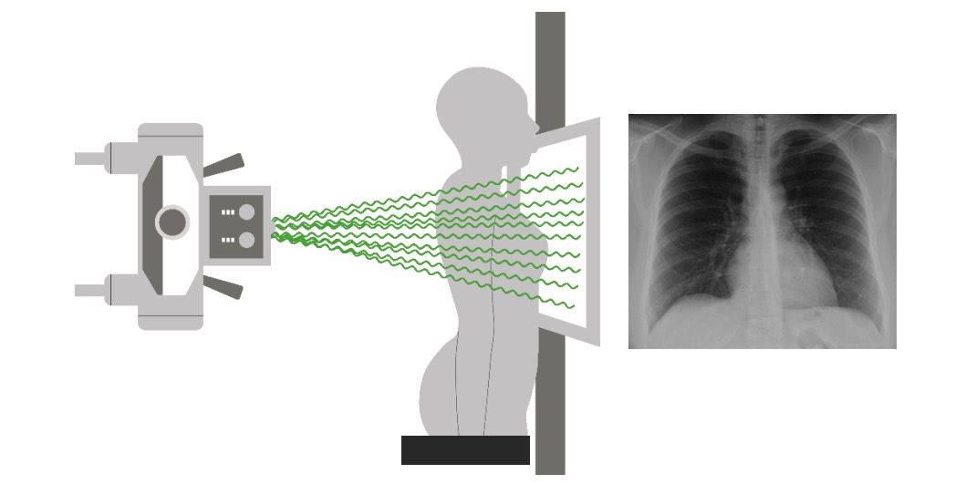
00:01 So let's take a look at the bony structures that can be evaluated on a radiograph. 00:06 You can see here both clavicles. 00:08 We often see clavicular fractures occur and sometimes their incidental findings. 00:13 So it's important to take a look at all of the bones carefully. 00:16 You can see the outline of the scapula which comes around to this way and projects posterior to a portion of the lung. 00:25 You can see liver in the upper abdomen and you really shouldn't see any pocket of air in this area. 00:34 Every once in a while you may see a little pocket of colon that can come up this way. 00:39 For the most part though, it really should just be in a soft tissue density, and if you do see air that's concerning for free air in the abdomen. 00:46 You can see the spinous processes as we saw earlier and they really should be midline within the trachea. 00:53 And then you should see air in the left upper abdomen and that usually represents stomach and possibly colon. 01:00 On the lateral view, you can have a good image of the thoracic spine, and this is a good view to take a look at for any kind of thoracic compression abnormalities or bony abnormalities. 01:13 So in this lecture, we've gone over multiple different radiographic projections of the chest, they include the PA, the lateral, as well as the AP, and decubitus. 01:25 We've also gone over different positions and how they change the appearance of the chest radiograph. 01:29 And we've gone over different types of techniques which you can also change the appearance of a radiograph. 01:34 We've also come up with a good algorithm and you should use this algorithm as you look at all the different chest x-rays that we'll be evaluating in these future talks.
About the Lecture
The lecture Bony Anatomy in Radiology by Hetal Verma, MD is from the course Thoracic Radiology.
Included Quiz Questions
Which of the following findings of a chest X-ray PA view is considered abnormal?
- Air space in the right upper abdomen
- Outline of the scapula
- Clavicles on both sides
- Spinous processes in the midline of the trachea
- Trachea in the midline
Customer reviews
5,0 of 5 stars
| 5 Stars |
|
1 |
| 4 Stars |
|
0 |
| 3 Stars |
|
0 |
| 2 Stars |
|
0 |
| 1 Star |
|
0 |
I recomiendes these vídeos because de review is wonderfull the radiologic anatomy is easy whit this method


