Playlist
Show Playlist
Hide Playlist
Scapula – Bones and Surface Anatomy of Upper Limb
-
Slides 01 UpperLimbAnatomy Pickering.pdf
-
Download Lecture Overview
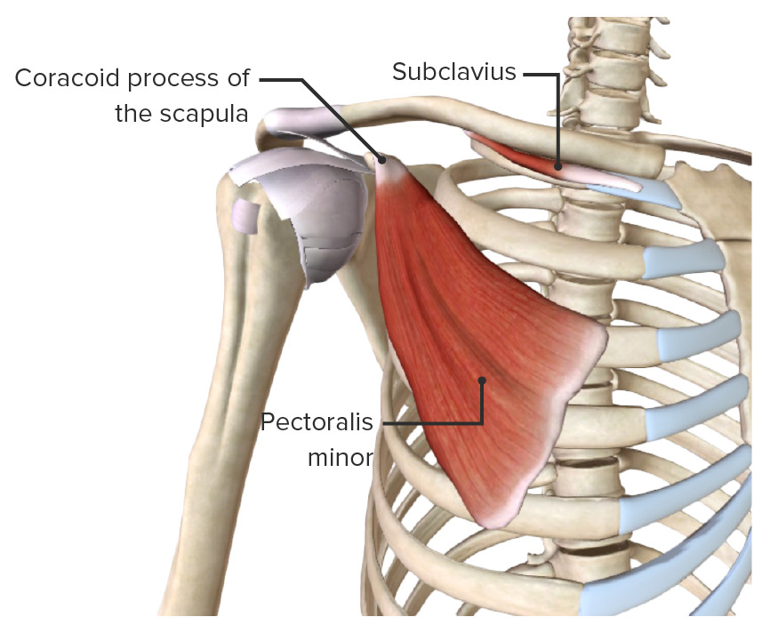
00:01 If we now move on to the scapula. The scapula is connected to the sternum by way of the clavicle. And we can see here we have numerous of views of a scapula. Here we've got a posterior view, here we've got a lateral view, the middle picture on this slide. And here we can see we have got an anterior view. We can see here on this side of the screen. So let's look at some features of the scapula. 00:30 As I mentioned it's a triangular, it's a flat bone and it sits on the posterolateral aspect of the thorax at the level of ribs 2-7. So it sits directly posterior to the rib cage at the level of ribs 2-7. The posterior surface which we can see here is divided by this prominent ridge. This is known as the spine of the scapula. We can see it dilates into this large structure here known as the acromion. But the spine separates the posterior surface of the scapula into a supraspinous. So supraspinous above the spine and infraspinous fossa. We have a supraspinous and infraspinous fossa and these fossae are important in offering muscle attachment that work on the shoulder joint and again we'll look at this in more detail. 01:27 So the posterior surface has this prominent spine, this ridge forming a supraspinous and infraspinous fossa. On the anterior surface it's quite featureless really but we just have a fossa again for subscapularis muscle and this fossa is known as the subscapular fossa which we can see here on this anterior surface. So the subscapularis muscle which we will cover in few lectures time is sitting directly between the ribs and the scapula. 02:04 So some more features. We have what are known as angles on the scapula. We have three angles. 02:12 We have a superior, an inferior and a lateral angle. So let's just orientate ourselves. 02:17 This is at the top on the posterior view. So we have a superior angle. The superior angle is connected to this inferior angle via this medial border. We can see this medial border of the scapula here. We have got a superior angle, this tip and we have got an inferior angle here. And these two are connected by way of this medial border. We also have a lateral angle here and that forms the three points of this triangle-shaped scapula. 02:49 The superior angle is connected to the lateral angle by way of this superior border and the lateral angle is connected to the inferior angle by way of this lateral border. So on the scapula, we have these three angles, superior, inferior and lateral. And these are connected via borders, medial, lateral and superior borders. And we can see these on the screen. 03:17 Superior angle, inferior angle, lateral angle. Medial border, lateral border, superior border. 03:27 And let’s have a look at a few more features that we can see. First of all, on this lateral border, we can see here, we have a neck of the scapula. So this narrowing where the superior border and the lateral border began to converge we have a neck and then we have a head, the head of the scapula. And this is characterized by a depression which is known as the glenoid cavity and that is going to articulate with the head of the humerus forming the shoulder joint of the glenohumeral joint. So we can see we have got the glenoid cavity here forming the head really and just medial to it we have the neck of the scapula along this lateral border. The superior border here we have an important feature and this is known as the suprascapular notch. The suprascapular notch is important as it allows the blood vessel to pass through over the top of the scapula and we will come to that in due course in some later lectures. So here we can see various features which we can see on this posterior view. We can see if we look at the anterior view we can again see we have got this neck of the scapula and we have got a head of the scapula forming the glenoid cavity and we can see the suprascapular notch. If we look at the spine, so let's just concentrate on the spine of the scapula there we can see starting medially from this medial border and as it travels laterally and it actually gets bigger and bigger until it forms this dilated bulge which is known as the acromion. And we can see this here on the posterior view. We can see it's prominently passing more laterally than the actual main body of the scapula. And we can see the acromion here on this anterior view. 05:17 And here on this lateral view, which as if you're looking from the lateral aspect, we can see we have got this costal surface. So this is the anterior surface sitting against the ribs and this is the posterior surface here, so it have skin along here. Here we can see the glenoid cavity and then radiating up here we have got the acromion. We can see it's almost running completely over the glenoid cavity and that is important as we come to realize because it can help to prevent superior dislocation of the humerus. So we have got the acromion and remember in a few slides previously we have the clavicular acromial ligament where the ligament attaching from the acromion to the clavicle. We will come back to that. So we have got the spine here of the scapula and that articulates with the clavicle. If we look at the head in a bit more detail then we have got the glenoid cavity. We can also see if we look clearly here with this lateral view, here is the glenoid cavity. We can also see if we look clearly here with this lateral view, here is the glenoid cavity. We can see we have got a supraglenoid and an infraglenoid tubercle. So small little elevations, it's all bony masses on the superior and inferior aspects of the glenoid cavity and these are important for muscle attachment as we will see both long head of biceps and triceps attached to the superior and infraglenoid tubercles respectively. So here we have got the glenoid cavity, and a supra and infraglenoid tubercles. 06:52 We will come back to these later on. The final feature I want to talk about is the coracoid process here and this is an important kind of bulge again coming up from the superior border and from the head of the scapula. And this is the coracoid process. Again it helps to form the muscle attachments we will talk about coracobrachialis in a few slides time and we can see here the coracoid process. Here again on the lateral view we can see coracoid process and here on this anterior view we can see again the coracoid process. So some important features here on the scapula.
About the Lecture
The lecture Scapula – Bones and Surface Anatomy of Upper Limb by James Pickering, PhD is from the course Upper Limb Anatomy [Archive].
Included Quiz Questions
Which of the following statements regarding scapula is correct? Select all that apply.
- The coracoid process is located on the lateral aspect of the superior border of the scapula.
- The glenoid cavity articulates with the clavicle.
- The glenohumeral joint is an articulation between the head of the humerus and a specialized region of the scapula.
- The posterior surface is the subscapular fossa.
At which thoracic rib level is the scapula located?
- 2–7
- 1–7
- 3–7
- 4–7
- 1–5
Which border connects the superior angle of the scapula with the inferior angle?
- Medial
- Lateral
- Superior
- Posterior
- Inferior
Which dislocation of the humerus does the acromion prevent?
- Superior
- Anterior
- Posterior
- Inferior
- Lateral
The fossa on the anterior surface of the scapula provides attachment to which muscle(s)?
- Subscapularis
- Supraspinatus
- Infraspinatus
- External intercostal muscles
- Brachioradialis
Customer reviews
4,5 of 5 stars
| 5 Stars |
|
1 |
| 4 Stars |
|
1 |
| 3 Stars |
|
0 |
| 2 Stars |
|
0 |
| 1 Star |
|
0 |
Helped me a TON! Great quick easy overview of the Scapular bone, and was much easier to learn and understnad throughout this video. Much better understandment than my antomy class.
I certainly liked the video format, where we can adjust the focus from the speaker to the slide, and use the cursor to aid in pinpointing the relevant aspects of the scapula. The only thing I found was missing was a little aside about the suprascapular notch, which was shown in the slide, but not alluded to in the actual video lecture!


