Playlist
Show Playlist
Hide Playlist
How to Read an Electrocardiogram (ECG): Introduction
-
Slides Learning to Read an ECG.pdf
-
Download Lecture Overview
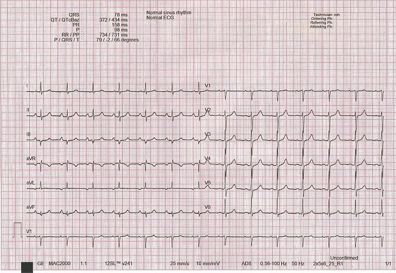
00:00 Hello. I'm Joseph Alpert, Professor of Medicine and Cardiology at the University of Arizona in Tucson, Arizona. I'm also the editor-in-chief of the American Journal of Medicine. This is the first of a series of 10 lectures that's going to give an introduction to how to read an electrocardiogram. The electrocardiogram is one of the most useful and simple diagnostic tests that we have as cardiologists and as internists and it can be mastered with a little bit of effort and a lot of practice. At the end of this lecture series, you will not be an expert in reading electrocardiogram, but you will understand and be able to read most of the fairly straightforward electrocardiograms. There will still be some subtle ones that are difficult and the answer to that of course is practice, practice, practice just like you were learning to play a musical instrument. You're not ready to perform in public after 10 lessons, but you will be quite familiar with the electrocardiogram following this series. So let's get started. First of all, it turns out that the human brain is particularly good at recognizing patterns and this is a genetic characteristic that was probably evolved millions and millions of years ago when you needed to recognize what was out there in front of you was something that might be able to be eaten or something that was looking to eat you. So, that's exactly what happens with the electrocardiogram. After a while when you become very expert, your brain allows you to recognize the pattern immediately when you see it. Now of course, there's going to be some steps where you're going to have to make some measurements but in fact you will immediately know "Oh I recognize that, that's a heart attack" and so forth. So I think after the end of this 10-lecture series, you will be able to do that and with practice you'll get very good at it. So, I'm sure you recognize immediately these 2 paintings. Right? On the left is van Gogh's starry sky, on the right is Leonardo da Vinci's Monalisa. How did you recognize them immediately? Because you know the pattern you've seen them so many times. In the end, I think you'll get to be able to read the electrocardiogram in a very similar way. You'll immediately recognize something and then you'll go back and do a little more detailed work on it. So when do the electrocardiogram start? It started in the early 20th century. A Dutch physician named Einthoven was the first to be able to record the electrical activity of the heart with any accuracy. Many have tried before, but he was the first that was successful. Now you can see from this very primitive early electrocardiogram, one had to have an arm in saltwater and a leg in saltwater and a huge apparatus in order to record the electrocardiogram. Well today, of course, with solid state instruments they're much more sophisticated, the amplification makes the images much clearer and now we use a very standardized protocol all over the world and the standards we arrived at a long time ago back in the 1930s by experts who came together to decide what would be the universal rules for an electrocardiogram. So, reading the electrocardiogram, as I said, it's like examining a fine artwork. First of all you get an overall impression something leaps out at you particularly if it's acute heart attack, acute myocardial infarction as we'll talk about. But then secondly, there has to be the more careful detailed analysis, things like heart rate and duration of the various intervals and we're going to go over that now. So, in the beginning, don't hurry, take your time, systematically check the heart rate, the timing of the intervals and the presence or absence of P-waves. So we're going to talk about what that means in a moment. Remember, many times you're going to be seeing an electrocardiogram it's been recorded by a computer and the computer will have read a diagnosis. It will give you some information. The computers usually write about heart rate and intervals, but it's often not right about the underlying rhythm and sometimes it even makes more serious mistakes. So every computerized electrocardiogram has to be overread by an experienced electrocardiographer. So the first thing you're going to look at is going to be the heart rate, that's the number of beats per minute. I'm going to give you a rule how you can calculate the heart rate from the EKG. Of course, it will already have been calculated for you if it's a computer-read ECG. Then we're going to do the various intervals. That's the various points on the electrocardiographic complex that you see on the right-hand side and we're going to go over how those intervals are derived and how long they are when it's normal. So let's start. The first wave in the electrocardiogram is the P-wave and this is the electrical signal made when the atria, that's the upper chambers depolarized. Now you're going to ask why P, Q, R, S, and T? Why not A, B, C, D, and E? Well, in fact in the early years where there were attempts in the early 20th century to try and record the electrocardiogram, there were many artifacts that were called A, B, C, and D and so forth all the way through the alphabet and finally Einthoven got to the letters P, Q, R, S, and T which were the accurate ones. So all the earlier ones were artifacts so that's why we use these terms. So here is a normal complex. You'll notice the P-wave, that's the electrical depolarization, the wave of electricity running down through the atria. 05:54 And then it arrives at the ventricle, that's the QRS, that big complex is the ventricle. 06:00 Ventricle has much more heart muscles so therefore it's a much bigger complex. And then you have something called the ST segment, which is a little pause before the ventricle resets itself and the resetting is the T wave and some patients will have the U wave, following the T wave. 06:18 This can be normal, it's absent in most electrocardiograms and it is a little more commonly seen when patients have low blood potassium so-called hypokalemia. So here again are the various segments, you'll notice that the so-called PR intervals is the segment that runs all the way from the beginning of the P wave to the QRS. It's divided into 2 segments, the duration of the P wave and then the PR segment that is from the end of the P wave to the beginning of the QRS. You then see in gray the QRS complex, that's the depolarization of both ventricles by the way, right and left although the left has a lot more muscle. So, the QRS is dominated by left ventricular muscle depolarization. There is then an ST segment where a little pause while the ventricle finishes the depolarization and gets ready to repolarize, that is to get ready for the next beat and that's during the T wave. The T wave is the repolarization wave and as I mentioned the U wave is often not present but maybe present as a very small wave following the T wave. The electrocardiogram is usually the first test done after the history and physical exam. It's often done in offices, it's very simple and many physician offices have their own system. Usually it's done by a technician in the office, it may be done by the office nurse. In the hospital, there' s a whole series of technicians who are on 24 hours a day to record accurate electrocardiograms. It is again the simplest, cheapest, and most easily obtained cardiovascular test. It has reasonable accuracy for a variety of heart conditions, for example arrhythmias or myocardial infarction or heart attack. However, it's not as accurate as determining the volume of heart muscle compared to an MRI, an echo, or CT scan. 08:18 But all of those are much more expensive, they require huge amounts of equipment and machinery, the electrocardiogram is really inexpensive and very simple and is almost always the first test done. One of the reasons it takes a long time to learn to read electrocardiogram is there are many nonspecific or nondiagnostic patterns and there are some differences between men and women and between different groups, for example, in the United States African Americans have slightly different normal values from European white males and so forth. So, all of these of course is in the computer and the computer is told before the EKG is taken what the patient's ethnic background is and so forth. So that helps in terms of the normals. And also, you'll get to recognize these things as you practice reading many electrocardiograms.
About the Lecture
The lecture How to Read an Electrocardiogram (ECG): Introduction by Joseph Alpert, MD is from the course Electrocardiogram (ECG) Interpretation. It contains the following chapters:
- Learning to Read an Electrocardiogram
- Reading ECGs
Included Quiz Questions
The QRS complex reflects which event?
- Ventricular depolarization
- Atrial depolarization
- Ventricular repolarization
- Time from start of atrial depolarization to start of ventricular polarization
- Junction between the end of ventricular depolarization and the start of the ST segment
The T wave corresponds to which of the following?
- Ventricular repolarization
- Atrial depolarization
- Atrial repolarization
- Junction between the end of the QRS complex and the start of the ST segment
- Time from the start of atrial depolarization to the start of ventricular polarization
The P wave corresponds to which of the following?
- Atrial depolarization
- Atrial repolarization
- Ventricular repolarization
- Junction between the end of the QRS complex and the start of the ST segment
- Time from the start of atrial depolarization to the start of ventricular polarization
Which of the following is true of the U wave?
- Commonly seen in hypokalemia.
- It is seen in hyperkalemia.
- It marks atrial depolarization.
- It is the junction between the end of the QRS and the T segments.
- It marks atrial repolarization.
Customer reviews
5,0 of 5 stars
| 5 Stars |
|
6 |
| 4 Stars |
|
0 |
| 3 Stars |
|
0 |
| 2 Stars |
|
0 |
| 1 Star |
|
0 |
Absolutely fantastic to explain the very basics of the cardiac cycle reflected in the ECG. THANK YOU!
so useful and great. Thanks a bunch for your efforts !!!!
Thanks a lot. Great effort Dr. Alpert. All the cardiology lectures are very useful.
Very lovely introduction, really like the analogy for reading ECG and and art - as a pattern recognition! Also reiterated bullet points, very comprehensive to start with! Thanks c:


