Playlist
Show Playlist
Hide Playlist
Trachea

-
Slides 03 Human Organ Systems Meyer.pdf
-
Download Lecture Overview
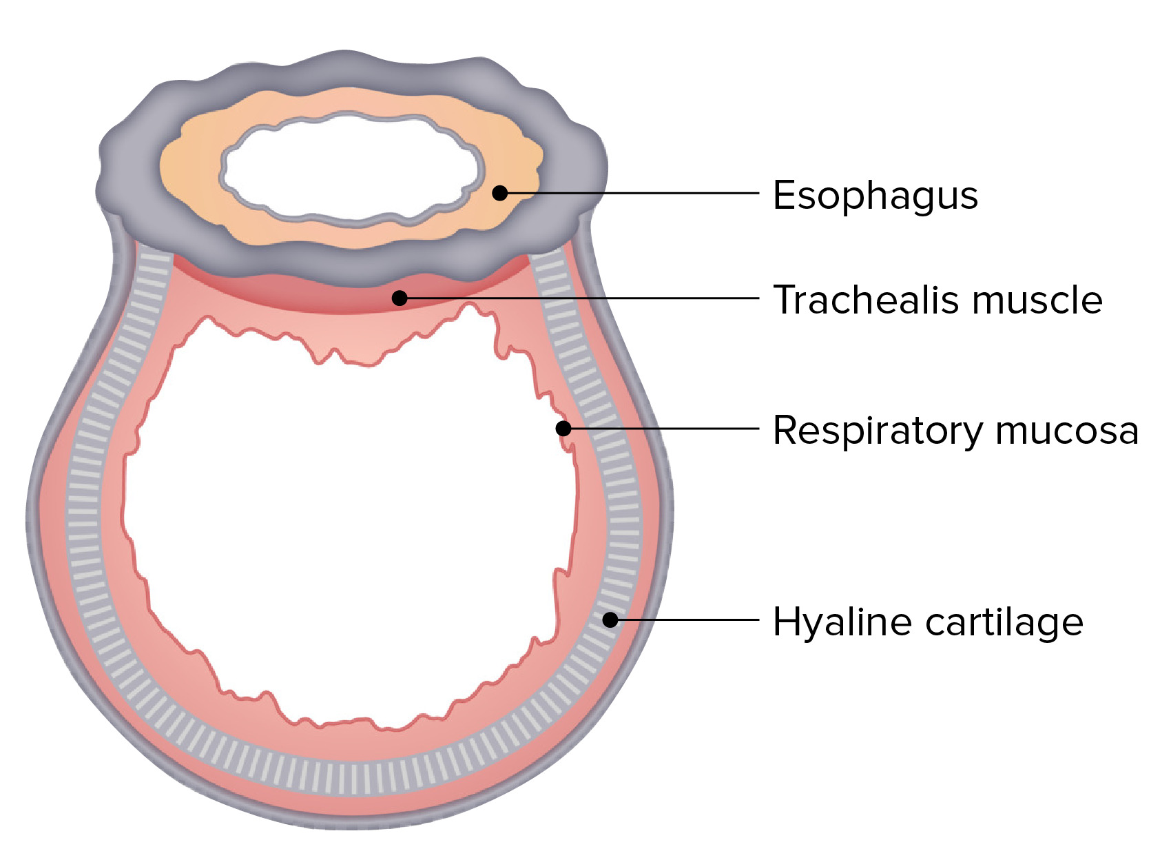
00:00 part of the swallowing process. Well let us look at the trachea. Trachea has got a number of components as I said they lie, we can feel them in our neck region, we have 16 to 20 of them, the rings of cartilage, horseshoe shaped cartilage and they joined at the ends of these horseshoes by some connective tissue and also by the trachealis muscle. The trachea has a mucosa again of respiratory tract epithelium. It has a submucosa to support that epithelium and also has, as I mentioned before, rings of hyalin cartilage and on the outside the adventitia, which is going to blend with surrounding structures. The trachealis muscle can alter the dimensions of the trachea and therefore, the amount of air flowing to the lungs and the tissue adjacent to the trachealis muscle then blends with the esophagus. The esophagus lies just behind the trachea. Now those are cartilage rings and they are separated from each other by connective tissue and also elastic tissue because the trachea alters its dimensions. 01:21 It can lengthen and contract during the process of air passing through it, down to the lungs, or out from the lungs. So there's elastic tissue in between these cartilage rings as well as the normal supporting connective tissue. Let us have a look at the epithelium of the trachea and components of the trachea in a little bit more detail. There's the elastic fibres sitting in the lamina propria. The elastic fibres are running in a longitudinal direction to the epithelium and these elastic fibres also give the lamina propria and the supporting tissues and the epithelium the ability to expand and lengthen and shorten along with trachea during the same thing that I've described a moment ago. The epithelium sits on a very prominent basement membrane and that basement membrane will support the epithelium and then just underneath the lamina propria, you find some submucosal glands. These are both mucous and serous and again they keep the surface of the epithelium moist. And again you maintain the mucous content on the epithelium to create that raft I mentioned earlier that can carry debris that is stuck to that epithelial surface, stuck to the mucous that can be carried up to where it can be swallowed or coughed and gotten rid off from the body because that epithelium is still ciliated and those cilia beat and move that mucous raft along. 03:06 The elastic fibres you see on the left-hand image actually divide. They are the division between the lamina propria, the very loose connective tissue supporting the epithelium just underneath the basement membrane and then below that there is the division between that lamina propria and the underlying submucosa. When you go down from the trachea towards the lung,
About the Lecture
The lecture Trachea by Geoffrey Meyer, PhD is from the course Respiratory Histology.
Included Quiz Questions
Which of the following statements regarding the trachea is MOST ACCURATE?
- It has 16-20 rings of hyaline cartilage.
- Its epithelium does not include mucus-secreting goblet cells.
- It is lined by pseudostratified squamous epithelium.
- It is located posterior to the esophagus.
Which of the following best exemplifies the shape of the rings of hyaline cartilage in the trachea?
- Horseshoe-shaped
- S-shaped
- Spiral shaped
- Longitudinal
Author of lecture Trachea

Geoffrey Meyer, PhD
Customer reviews
5,0 of 5 stars
| 5 Stars |
|
1 |
| 4 Stars |
|
0 |
| 3 Stars |
|
0 |
| 2 Stars |
|
0 |
| 1 Star |
|
0 |
eloquent and calm voice which is easy to listen and understand. its great to have lectures which are easy listening like this. not too fast paced to follow and also not to slow that i fall asleep. thank you .
