Playlist
Show Playlist
Hide Playlist
Chest X-ray – Diagnostic Imaging
-
Slides DiagnosticImaging RespiratoryPathology.pdf
-
Download Lecture Overview
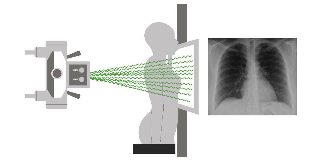
00:01 Students are always asking me about imaging. 00:04 Well, you know that imaging is a huge part of your medical education and that’s for as when you need to know it, you don’t wait until your specialization to get into it. 00:15 It’s immediately during medical school and the more that you know about what you’re looking for with imaging the better off you’ll be. 00:22 Let’s get started. 00:24 Take a look at a normal chest x-ray. 00:26 On a normal chest x-ray you’ll notice the following. 00:29 If you take a look at the areas of the lungs that are lucent but there’s just enough opacity in which there’s vasculature that you should be seeing. 00:40 Now, what we’ll do is as we go into emphysema or perhaps even a pneumothorax is that, well, with emphysema the both areas of the lungs are completely lucent and they then appear completely black. 00:56 That is not normal, that is a pathology. 00:59 Or in pneumothorax when you have one side of the lung, say for example it is a spontaneous type of pneumothorax where it then collapses then you would have one area of the lung which is completely black but you’ll notice here that there should be some opacity then representing vasculature and also on your left side you should be able to find the heart. 01:22 And with that heart being present there, understand that this is not going to be laterally displaced so this is not cardiomegaly. 01:30 You have just enough of your heart in which you see a cardiac silhouette then coming out towards the left representing the apex at the midclavicular line. 01:43 Take a look at the left side, go to midclavicular approximately 5th intercostal, you can scrape down and that then represents your apex. 01:51 These things you keep in mind because if you get an x-ray and has cardiomegaly that apex will be a lot more displaced. 01:58 Now, the type of x-ray that you’re seeing on the right will then be your AP diameter, anterior/posterior, and something like this would be completely pathologic if we see a condition such as emphysema where the lung itself is completely widened or filled or hyperinflated.
About the Lecture
The lecture Chest X-ray – Diagnostic Imaging by Carlo Raj, MD is from the course Pulmonary Diagnostics.
Included Quiz Questions
Which of the following findings is considered normal in a chest X-ray?
- Lungs are lucent, but there is just enough opacity that is represented by the vasculature.
- Lungs are opaque, but there is just enough translucency that is represented by the vasculature.
- Lungs are lucent, but there is just enough opacity that is represented by the nerves.
- Lungs are opaque, but there is just enough translucency that is represented by the nerves.
- Lungs are opaque.
On a chest X-ray, the right side of the lung field looks hyperlucent and the other side looks lucent with opacity represented by the vasculature. What is the most likely diagnosis?
- Right-sided pneumothorax
- Left-sided pneumothorax
- Right-sided pleural effusion
- Left-sided emphysema
- Right-sided pneumonia
Which of the following statements is true regarding the cardiac silhouette on a normal chest X-ray?
- The apex is seen on the left, at the mid-clavicular line, approximately at the level of the 5th intercostal space.
- The apex is seen on the right, at the mid-clavicular line, approximately at the level of the 5th intercostal space.
- The apex is seen on the left, at the mid-axillary line, approximately at the level of the 5th intercostal space.
- The apex is seen on the left, at the mid-clavicular line, approximately at the level of the 6th intercostal space.
- The apex is seen on the left, at the mid-clavicular line, approximately at the level of the 4th intercostal space.
Customer reviews
5,0 of 5 stars
| 5 Stars |
|
5 |
| 4 Stars |
|
0 |
| 3 Stars |
|
0 |
| 2 Stars |
|
0 |
| 1 Star |
|
0 |


