Playlist
Show Playlist
Hide Playlist
Introduction – Bones and Surface Anatomy of Upper Limb

-
Slides 01 UpperLimbAnatomy Pickering.pdf
-
Download Lecture Overview
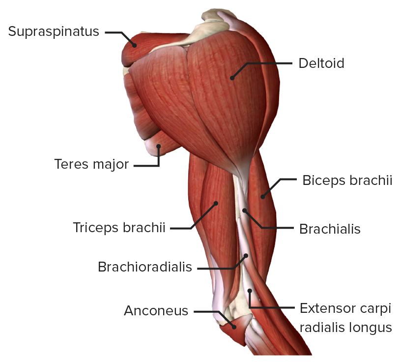
00:01 In this lecture, the first on the series about the upper limb, we are going to look at the surface anatomy and osteology of the entire upper limb. So we are going to look at the surface anatomy of the shoulder, the arm, the forearm and the hand, all of these regions making up the upper limb. And then we are going to move onto the osteology, we'll look at the clavicle, the scapula, the humerus, ulna, radius, the carpal bones, the metacarpals and the phalanges and in each of these bony structures, we'll look at specific landmarks and these are important for muscle attachments. We'll then finish by looking at various movements that are performed by the upper limb which is a complex part of the appendicular skeleton enabling a great range of movements. So if we just look at the general arrangement of the upper limb, then on this slide we can see we have a nice body plan of both the male here and the female. And on the male, we can see indicate here we have got the upper limb, specifically we have got the arm region here which is connected to the trunk by way of the shoulder which we will talk more about here and then connecting the arm to the forearm we have the elbow joints which we can see here and then most distally furthest away from the main core of the body we find we have the hand. The hand member connected to the forearm via the wrist joint. On this picture here, we can see an anterior view of the upper limb, we can see more features more surface features. We can see we have this shoulder region and we can see a region known as the axilla. This is your armpit. 01:52 And we can see we have the deltoid. This muscular region that covers the shoulder joint and we'll look at the deltoid muscle in quite some detail as that covers the shoulder joint. 02:03 We then pass down distally towards the elbow and between the elbow and the shoulder we find we have the arm. We can see a groove here that is separating biceps brachii and triceps brachii here and this we can see the nice cleft this way you can palpate the brachial artery. So there are some important surface landmarks here. 02:26 We can then move on to the elbow joint and we have a prominent bulge on the medial aspect of the elbow joint. This is known as the medial epicondyle and that is important because the ulnar nerve runs alongside this structure and we will cover these details throughout this lecture series. We then pass distally from the elbow joint and we see the forearm, we can see the anterior region of the forearm. We have a prominent bulge on the lateral surface of the forearm and this is due to a really important muscle known as brachioradialis. 03:02 So we can see a bulge here caused by brachioradialis. We can then move distally again and we can see the hand. The hand is connected to the forearm via the wrist joint where the carpal bones connect to the radius and the ulnar to form this wrist joint. And we can see we have the palm of the hand, and then we can see we have the thumb, the index finger, the middle, the ring and the little finger that make up the digits of the hand. So these some key surface landmarks we can see on the anterior aspect of the upper limb. 03:39 If we look at the posterior aspect, then again we can see we have this muscular region here caused by the mass of deltoid muscle. And then we can see the posterior region, we can see some impressions for the various heads of triceps muscle. Again we can see the medial epicondyle, this time from the posterior view just as we saw it on the anterior view here. 04:04 We can see the olecranon, this bony prominence on the posterior surface of the elbow. 04:09 Once again if we move into the forearm we can see we have this posterior aspect here and again on the lateral aspect, running along the same aspect as the thumb this lateral aspect here. We can see we have brachioradialis. If we pass to the hand once again we can see the thumb, index, middle, ring and little fingers again that make up the digits and we can see this dorsum of the hand. Remember that forearm is connected to the hand via the wrist joint and we have two what are known as styloid processes of the radius and the ulna. And we'll look at this in detail when we look at the osteology. But both these styloid processes of the radius and the ulna, the radius being lateral, the ulna being medial. 05:00 Then we can see these bony prominences and these can be palpated at the wrist joint. 05:07 So this region that we can see here on the slide is the part of the superior appendicular skeleton and it forms the upper limb. The upper limb is important because it has a high level of mobility. A high level of mobility which is given to it via the pectoral girdle which we will see in a few slides time. It is also characterized by its ability to grasp structures. The hand is a really important structure at the distal end of the upper limb and its ability to manipulate the fingers, to hold or to grasp structures. So when you are holding a pencil to write, it is a complex arrangement of movements that allows your fingers to assume this position. It is the key difference from the lower limb. 05:54 The lower limb doesn't have this much mobility as the upper limb and also it doesn't have this much function of the feet comparted to the hand. So it is lot easier to pick up structures with your hands than it's with your feet. However the main benefits of the upper limb being its mobility, isn't the same as the lower limb because it doesn't need to be as mobile and the lower limb is important for stability. So you don’t have a greater range of movements, but it is important in being able to make sure your posture is correct. 06:26 Being able to support the entire weight of your trunk. So there is a battle between the upper limb and the lower limb. The Upper limb increasing mobility but it has got a high level of instability. So it is more prone to having dislocated shoulder joints. 06:48 There is a whole series of specialized joints in the upper limb. I have mentioned one of them the shoulder joint and we will see that in this lecture series. But the elbow joints and various joints most distally are very specialized allowing for this increased range of movements. As I mentioned also the axial skeleton is connected to this appendicular skeleton via the pectoral girdle and we can see that in the next few slides.
About the Lecture
The lecture Introduction – Bones and Surface Anatomy of Upper Limb by James Pickering, PhD is from the course Upper Limb Anatomy [Archive].
Included Quiz Questions
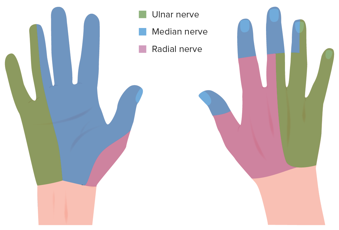
Which statement is true about the ulnar nerve?
- It runs posterior to the medial epicondyle.
- It runs posterior to the lateral epicondyle.
- It runs anterior to the lateral epicondyle.
- It runs anterior to the medial epicondyle.
- It is not related to the elbow joint.
Which bone is the lateral bone of the forearm?
- Radius
- Humerus
- Ulna
- Tibia
- Fibula
Author of lecture Introduction – Bones and Surface Anatomy of Upper Limb

James Pickering, PhD
Customer reviews
4,7 of 5 stars
| 5 Stars |
|
2 |
| 4 Stars |
|
1 |
| 3 Stars |
|
0 |
| 2 Stars |
|
0 |
| 1 Star |
|
0 |
Straightforward and extremely well organized. I like the way he relates parts of anatomy to one another.
simple, understandable, comprehensive . im looking forward into details on the anatomical structures of the limbs
easy to understand.... superb mcqs awesome lecture videos which helps to understand the concept better..
