Playlist
Show Playlist
Hide Playlist
Pulmonary Clinical Anatomy: Upper Portion


-
Slides OverviewOnDyspnea RespiratoryPathology.pdf
-
Download Lecture Overview
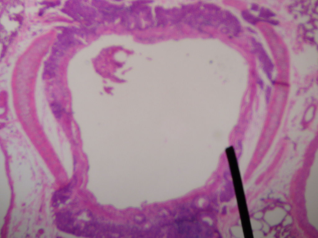
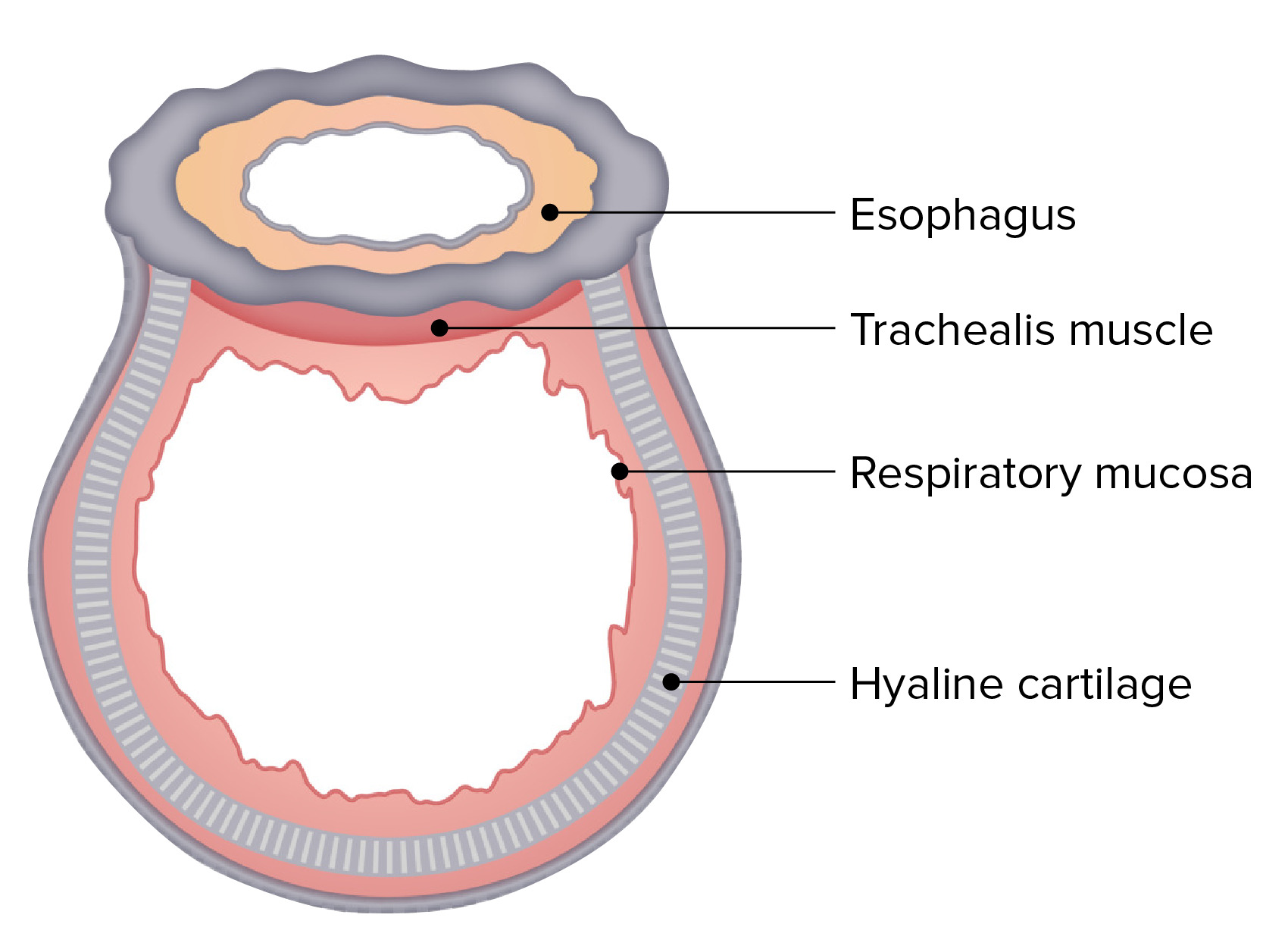
00:01 At first, we will take a look at the upper portion and this is your tracheas, it's cartilaginous. And it has mucociliary clearance which means that it is made up of columnar. 00:08 Let me give you another example. Say that you are smoking, and if you are smoking then what has to happen. Well, the trachea and company have to protect themselves, don’t they? And how do you protect yourself against smoke? Not with columnar cells. It's squamous cells. So, if the columnar cell has to turn into squamous cell, what do you call this? Good. Metaplasia. You see the point here. As we move forward and we go deeper beyond the bronchi into the bronchiole, then we get into goblet cells and it will produce quite a bit of mucous. And what does mucous mean to you? For example, let's say that you have three month of productive cough for two consecutive years. That is the definition of what? Chronic bronchitis. So at this point, as we move forward, the terminology becomes very important. It is important that you remain alert at all the times so that you understand my language and my terminology so that you know as to what I am referring to, kind of like what I have been doing in my entire course. 01:07 Okay, now, what’s beautiful about this is the fact that well, take a look at your right first. Proximal portion of the pulmonary tree. There is my trachea, and there is your bronchi. Now, we are going to put in some pathologies. We said that as far as the upper structure is concerned that it is made up of cartilage. Has to be, because you want that to be supported. You want that to strictly conduct air without any interference and down into alveoli. So why would you ever want premature collapse? You don’t. You need those cartilaginous rings. Now, what you are seeing here in blue will be the infections and what you are seeing here in red will be the diseases. Let's take a look at the parainfluenza virus. 01:47 You have heard of laryngotracheal bronchitis. Now, close your eyes and think of this anatomically in terms of sequence. What’s the first structure? Larynx. What is the next structure, distal? trachea. What’s after that? Bronchi. There you have it. So, instead of memorizing this, while you should already know from anatomy, the sequence. And so what am I referring to? What is this? It is parainfluenza. This is going to be your croup, isn’t it? So, your croup has your type of cough to it, but here, because how proximal it is. Now, if you know what a steeple is on top of a building such as a church then it is going to be that area of the building or the roof where it comes together like a steeple. Well, in parainfluenza or your croup, one of the most common causes of croup is parainfluenza. So, if you want C-R-O-U-P-P-P, often times the “P” is silent, but anyhow. Point is, it is a steeple sign that you might find on x-ray where, look where you are, up in the proximal portion. 02:50 And we have another infection number 5 and well take a look at this, located blue, circle 5 and that’s your bronchopneumonia. Allow the name to speak to you. “Broncho” meaning to say that you have involvement of the bronchi. So, we will see this later on as well as we get into distal segments, but giving you an understanding that this is involving the bronchi. A common organism here would be Klebsiella pneumonia. When you think about Klebsiella pneumonia, you should be thinking about perhaps this, you mean, if you have fun with it a little bit. Elderly patient and maybe perhaps a nursing home and they are hiding a bottle of alcohol. And every so often, they take to the canter and take a swig. And so, therefore, common pneumonia that you might find in elderly/perhaps alcoholics, kind of put them together there, if that helps you, would be the organism Klebsiella pneumonia. In microbiology, you have gone through different pneumonias and what they look like. For example, staph aureus, would be, well, what's aureo, what’s AU mean to you on the periodic table? Gold, isn’t it? So therefore, the type of phlegm or the productive cough that you have would be yellow, goldish clear. Say that it's something like pseudomonas. Well, that begins with a “P”. There is a little microbiologic agent called pyocyanine. What color is your sputum there? Green. Often times, pseudomonas as well, rarely, but still could result in your nails becoming green. Pyocyanine is an interesting component of Pseudomonas. Those would be infections, diseases. 04:30 What are squamous cell carcinoma or small cell? Now, the reason that I have put both of these together is because when we get into x-ray, we talk about lung cancers. While these cancers will be located centrally, and by that I mean it will be located by the mediastinum. 04:45 Is that understood? Now squamous, normally speaking, if you remember the trachea and the bronchi, then as you said earlier, you will undergo the process of what physiologic adaptation? Good, it is called metaplasia. Let me ask you something. What if you are smoking and your patient unfortunately develops progressive dysphagia? What's dysphagia mean? Difficulty with swallowing. That would be in esophagus. Be careful. Now, smoking obviously could result in cancers up and down the body, but I have given you two here. One would be in the trachea, and this would be something like, maybe squamous cell and that is a metaplastic transformation. 05:25 But, what if you were in the esophagus? Is that a metaplastic transformation before you went into cancer, before you went into dysplasia and then cancer? No, it wasn’t. Is that clear? Really? Because esophagus made up of what kind of histology, normally? Think. 05:42 Non-keratinized type of, you tell me. Good, squamous. So, it began as squamous. 05:48 Your smoking ended up as squamous cancer. So, the process never went through a metaplastic change before going to dysplasia and cancer. Is that clear? But, Dr Raj, I thought that you had Barrett’s esophagus? Yes, Barrett’s esophagus is due to why? Oh, reflux of acid. 06:05 Okay, so, what we wished to do here as we go through with this is make sure that you are clear about definitions, common things that you'll encounter on your board exams and things that you want to look out for over and over and over again. 06:17 We are strictly in the trachea. Now, small cell, extremely aggressive, another name for small cell, you should know is Oat cell. Which you notice isn’t here is adenocarcinoma, which is the most common lung cancer. All these three will group together later and it is called bronchogenic. 06:34 Now, you take a look at number 3. Number 3 is chronic bronchitis. Where are you? Bronchi. Proximal portion. What's happening. You might have heard of something like a Reid index. If you have, it will make perfect sense to you. Chronic bronchitis, inflammation of the bronchi, that is taking place you are producing quite a bit of mucous, usually due to smoking. And the way they will approach obstructive diseases in the lung will be rather novel for you and it will be much simpler and it's current day practice that you have to be familiar with because chronic bronchitis, emphysema and asthma and I will show this as we go through with this, has overlapping type of signs and symptoms, as you shall see. You will have fun with this and the definition of chronic bronchitis is three months of consecutive productive cough over two consecutive years. You must have a span of three months in which your patient is coughing for three consecutive months, keep that in mind, because you will be given something like that at some point in time in terms of history on your clinical rotations, on exams and so forth. 07:46 Move on.
About the Lecture
The lecture Pulmonary Clinical Anatomy: Upper Portion by Carlo Raj, MD is from the course Introduction to Pulmonary Pathology.
Included Quiz Questions
Which of the following transformations must occur to produce metaplasia of the trachea due to smoking?
- Columnar cells to squamous cells
- Ciliated columnar cells to non-ciliated columnar cells
- Columnar cells to cuboidal cells
- Ciliated columnar cells to ciliated cuboidal cells
- Columnar cells to pseudostratified columnar cells
Which of the following is FALSE regarding laryngotracheobronchitis?
- It can lead to chronic bronchitis.
- It is commonly referred to as croup.
- It is often caused by the parainfluenza virus.
- It displays the characteristic "steeple sign" on the X-ray.
- It affects the proximal conducting portion of the lung.
Which of the following is the most common causative agent of pneumonia in elderly, alcoholic patients?
- K. pneumoniae
- S. aureus
- P. aeruginosa
- Parainfluenza virus
Which of the following are required to make the diagnosis of chronic bronchitis?
- At least 3 months of productive cough per year, for at least 2 consecutive years
- At least 6 months of productive cough for at least 2 years
- At least 3 months of productive cough for at least 3 years
- At least 6 months of productive cough for 3 years
- At least 3 months of productive cough for 5 years
Author of lecture Pulmonary Clinical Anatomy: Upper Portion

Carlo Raj, MD
Customer reviews
5,0 of 5 stars
| 5 Stars |
|
1 |
| 4 Stars |
|
0 |
| 3 Stars |
|
0 |
| 2 Stars |
|
0 |
| 1 Star |
|
0 |
Love the explanation here about diseases and infections! Will never forget why squamous and small cell carcinomas lie medially now! I also really enjoy the interactive component of your lectures! Keeps me alert even after many hours of study Thanks Dr Raj
