Playlist
Show Playlist
Hide Playlist
Trisomy 21

-
Slides Trisomy 21.pdf
-
Download Lecture Overview
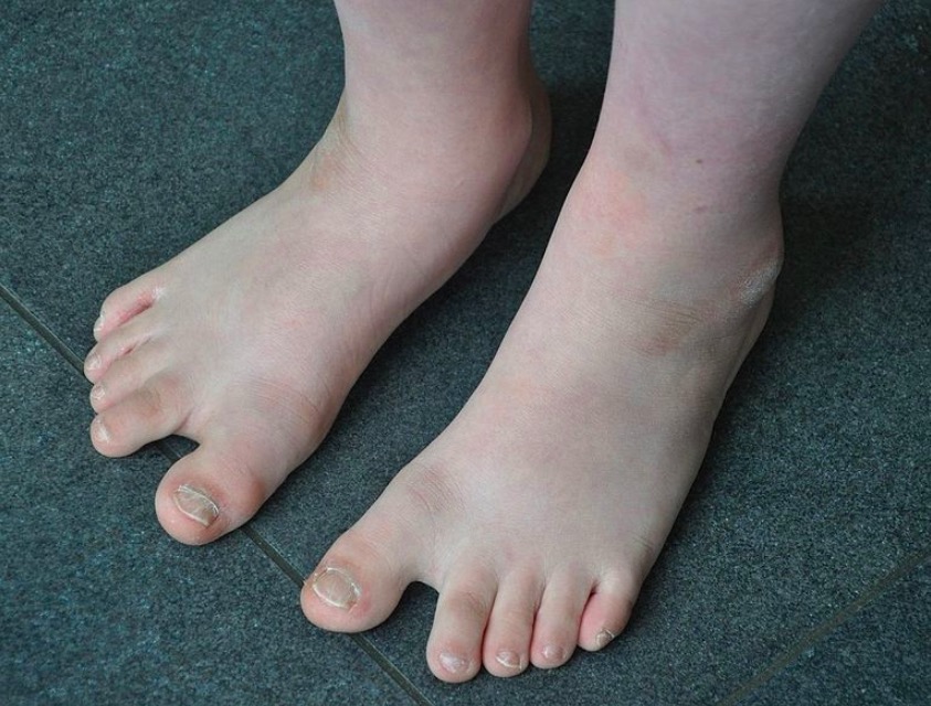
00:01 In this lecture, we will discuss Trisomy 21 or Down syndrome. 00:05 Trisomy 21 is when a patient has 3 copies of their 21st chromosome in every cell in their body. 00:13 These patients have characteristic facial features like you can see here. 00:17 They have hypotonia, they have intellectual disability and they may have congenital malformations and we?ll go through of this more carefully in this lecture. 00:28 So how is this happened? The most common times about 94% at a time, this is from non-disjunction. 00:37 Basically, one parent creates two normal gametes and the other parent creates one gamete with two copies and the other one simply isn?t viable. 00:47 Then through freak occurrence, the gametes with two copies fertilizes a normal egg or the egg has two copies and is fertilize by a normal sperm and this results in a patient with 3 copies of the 21st chromosome. 01:02 So, this is non-disjunction, this is about 94% at a time. 01:06 About 6% of a time this will happen through Robertsonian translocation. 01:13 Robertsonian translocation is a little bit complicated so I wanna walk you through at this one time so we are all in the same page. 01:21 We?ll repeat Robertsonian translocation when we talk about other trisomies. 01:25 But here we have a patient who is normal and a spouse who has a Robertsonian translocation, this spouse is completely normal from the standpoint of how they appear to you or how they interact with you. 01:37 This patient has two blue chromosomes 14, I just chose 14 as an example. 01:44 Then for chromosome 21, a piece of it has been translocated onto the end of one of those 14 chromosomes. 01:52 So, they have the appropriate amount of chromosomal material in each cell, it?s just mislocated with half of one chromosome being adhered to another. 02:03 That?s the Robertsonian translocation, but now let?s watch what happened when a patient who has translocation then goes and has a child with a normal individual and what happens to the various opportunities for their offspring. 02:18 So, let?s consider a case where a parent who has a normal genome is going to have a baby with a parent who has a Robertsonian translocation. 02:27 The normal parent will make two gametes, these will both be normal. 02:31 One copy of 21 which are the red ones and one copy of 14 which are the blue ones. 02:37 However, the parent who has translocation carrier will make 6 possible gametes and you can see them all drawn here with the lowest one being a normal gamete, but the others all being variations. 02:51 Now, when this two parents make an offspring, it?s possible that they may make a normal offspring as you can see in this picture with the lowest gamete and a translocated carrier matching up with one of the normal gametes from the normal patient. 03:07 However, they may also make Trisomy 14 that?s not viable. 03:13 That child will not come to be or they could create yet another translocation carrier like the translocation carrier in the first example. 03:24 This child will grow to be a carrier of Down syndrome, but will not have any manifestation of Down syndrome or they could make a monosomy 21, which is not viable. 03:36 This child will not become a being or alternatively, they could create a translocation such that this patient has Down syndrome. 03:46 They have essentially 3 copies of the 21st chromosome, one of them is adhered to there 14th chromosome or alternatively, they could create monosomy 14 that?s not viable either. 04:00 So essentially, the odds are for this couple that they can either have a normal child. 04:06 A child who is put at risks for creating children in the next generation in a similar way or a child with Down syndrome. 04:15 So, let?s look now at risks for Down syndrome. 04:19 The biggest risk that we have to consider is the maternal age with women who get older. 04:27 As they get older, it?s more and more likely for them to create eggs that have an extra copy of a 21st chromosomes and you can see the risks here. 04:36 So generally, we consider 35, a point at which the risk starts to really come up and after 40 it comes up dramatically and those are the risks as you can see. 04:49 So, let?s say we have a baby and we're wondering whether they might have Down syndrome. 04:55 This is a diagnosis that frequently made on physical exam and there are some classic physical exam findings that we can see. 05:01 One is they can have epicanthal folds or upslanting fissures of their eyes. 05:07 They may have midface hypoplasia, you can see that a little bit in this child. 05:12 They may have brachycephaly, they may have a little bit of excessive skin behind the neck, sometimes we can see that in utero on ultrasound. 05:21 You may see something called a palmar crease, this is where that crease goes straight across the hand instead of branching like it normally does. 05:30 You may see a slight gap between the first and second toes much like a flip flop sandal, it's where that flip flop post would go and you may see that gap there and you can see small white spots in the iris which are called Brushfield spots that can be associated with Down syndrome. 05:50 Patients with Down syndrome have other issues that can be more consequential, one examples congenital heart disease and this can show up at as many as 50% of patients, and the most common is what we called an endocardial cushion defect which is also called the common AV canal. 06:06 This is when they have both of VSD and ASD essentially creating one large chamber in the heart. 06:13 Patients with Down syndrome are increased risk for duodenal atresia about 12% at a time. 06:19 As you can see here we?ll have that classic finding of the double bubble sign on abdominal x-ray very early in life that represents quite quickly with inability to eat and inability to defecate and you can see that double bubble area where one bubble is in the stomach and the other bubble is the query of air before that area of duodenal atresia. 06:39 Patients may develop congenital hypothyroidism, this hopefully will be picked up on the new born screen. 06:45 If not, we want to address it as quickly as possible. 06:49 These infants happen to have it by 1% at a time among patients with Down syndrome and hearing loss and especially, frequent otitis media is common in patients with Down syndrome as well. 07:01 They may develop eye disease such as cataracts or refractive errors. 07:06 They may develop poor growth. 07:08 In fact, patients with Down syndrome should be plotted along their own special growth curve which is available online. 07:15 They may have increased risk for celiac disease, they may have atlantoaxial instability. 07:22 So, atlantoaxial instability is instability of C1 on C2 of their vertebrae, we used to recommend x-ray screening. 07:33 Though, now we?ve decided that is no longer particularly beneficial. 07:36 Although it may be required for participation in the special olympics which is important for these kids. 07:43 Additionally, they may have an increased risk of Leukemia and that is very important.
About the Lecture
The lecture Trisomy 21 by Brian Alverson, MD is from the course Pediatric Genetics.
Included Quiz Questions
Which of the following is not a typical finding in trisomy 21?
- Nail pitting
- White spots in the iris
- Space between 1ˢᵗ and 2ⁿᵈ toe
- Linear crease on palm
- Epicanthal folds
Which of the following is a major risk factor for Down syndrome?
- Advanced maternal age
- Paternal diabetes
- Paternal hypertension
- Male sex
- Large for gestational age
Which of the following is not a feature of Down syndrome?
- Skin deficiency behind the neck
- Epicanthal folds, upslanting fissures
- Transverse palmar crease
- Sandal gap
- Brushfield spots
Which of the following is the most common pattern of inheritance of Down syndrome?
- Nondisjunction
- Reciprocal translocation
- Deletion
- Missense mutation
- Robertsonian translocation
Which of the following is not associated with Down syndrome?
- Wilson disease
- Endocardial cushion defect
- Hypothyroidism
- Hearing loss
- Duodenal atresia
Which of the following may lead to cervical spine compression and is seen more commonly in patients with Down syndrome?
- Atlantoaxial instability
- Hypothyroidism
- Vertebral fracture
- Increased cervical lordosis
- Accessory cervical rib
Author of lecture Trisomy 21

Brian Alverson, MD
Customer reviews
5,0 of 5 stars
| 5 Stars |
|
1 |
| 4 Stars |
|
0 |
| 3 Stars |
|
0 |
| 2 Stars |
|
0 |
| 1 Star |
|
0 |
Really enjoy Dr. Alverson's approach to teaching. I like how he goes into important details and remains enthusiastic and clear with his explanations.
