Playlist
Show Playlist
Hide Playlist
Pediatric Choledochal Cysts

-
Slides Conjugated Hyperbilirubinemia and Liver Diseases Pediatrics.pdf
-
Download Lecture Overview
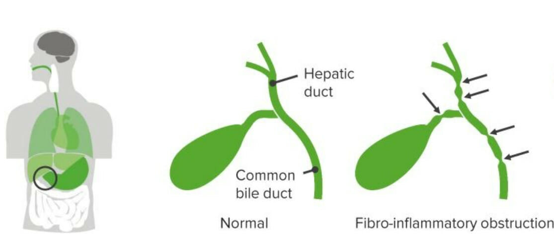
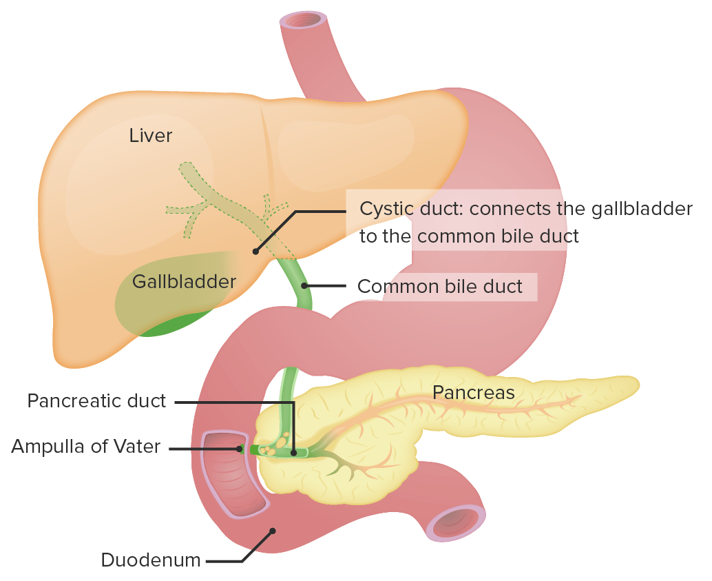
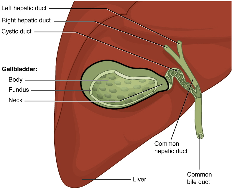
00:02 Let’s shift gear and talk a little bit about choledochal cysts. 00:06 Choledochal cysts are a congenital abnormality that cause abnormal enlargement of the bile duct. 00:13 This causes obstruction and hepatic congestion and eventually liver failure. 00:20 It’s not that uncommon. 00:22 You can see here a picture of a patient with a gallbladder and a large excised choledochal cyst. 00:31 It happens about 1:100,000 to 1:150,000 patients. 00:36 So you’ll see one every several years in a large birthing institution. 00:42 There are several different types of cysts. 00:44 I don’t think it’s likely that the test is going to quiz you on which type of cyst it is but it is important to remember that there are different types, and so elucidating what the type is may be beneficial in terms of understanding exactly what’s going on with that patient. 01:00 By far the most common is a cystic dilatation of the extrahepatic biliary duct, which you can see here. 01:09 You can here on your slide is a normal biliary tree and then next to it, in that common bile duct, there’s just an outpouching or cyst that’s formed along that duct. 01:22 Additionally, patients can have an abnormal pouch or a sac opening from the duct, this is a type 2. 01:29 A type 3 is a cyst, which is located within the wall of the actual duodenum. 01:35 A type 4 is when you have swellings both in the intrahepatic and extrahepatic biliary tracts, so it’s inside the liver and just outside it. 01:46 And type 5 is when there are multiple intrahepatic cysts. 01:50 This is the least common type. 01:54 So key symptoms for any child with choledochal cyst is typically they will have right upper quadrant pain. 02:01 Now, that’s on the test. 02:02 It’s sometimes hard to truly determine where the quadrant pain is in an infant, but you should see it. 02:08 But key -- key and highly likely to show up on your test -- these children will have jaundice and they will have acholic stools. 02:17 So they’ll have acholic stools again because that bile isn’t getting into the stools and their jaundice will be from the hyperbilirubinemia, which is conjugated. 02:27 Often, they may have fever or infection as a result of these cysts becoming infected. 02:33 They may develop pancreatitis and they often will have a palpable mass in the right upper quadrant. 02:40 You may be able to palpate this cyst. 02:44 In general, we’ll get some labs. 02:46 We’ll get bilirubin, which should be elevated and should have a high conjugated hyperbilirubinemia. 02:52 These patients will often have elevated alk phos, AST, ALT, and a high GGT as well because again the biliary tree is involved. 03:02 As in other things, we’ll get a coagulation profile, a PT/PTT, to assess the function of the liver and see how badly off they’re doing. 03:12 And a CBC is often obtained as well. 03:16 So the definitive diagnosis though is made through an abdominal ultrasound. 03:20 These cysts show up very nicely on abdominal ultrasound. 03:24 Rarely, we may get abdominal CT, maybe an MRI instead. 03:30 The MRI has the risk of needing sedation. 03:33 The CT is a little bit quicker, but has the risk of radiation. 03:38 Most centers are moving more towards ultrasound. 03:41 However, sometimes the surgeons want a really good idea of what kind of choledochal cyst this is that they’re dealing with and so sometimes we’ll get a magnetic resonance choleangiopancreatography or an MRCP to try and get a sense of exactly what is the structure, which type is this, so the surgeons have an idea of what they’re getting into. 04:04 Treatment of the choledochal cyst is a complete excision and a construction of a biliary enteric anastomosis. 04:12 Sometimes it’s Kasai procedure. 04:14 It really depends on what the problem is and this is what the surgeons are going to look into after the imaging is done. 04:22 So that is an assessment of the major kinds of causes of conjugated hyperbilirubinemia in young children. 04:31 Thanks for your time.
About the Lecture
The lecture Pediatric Choledochal Cysts by Brian Alverson, MD is from the course Pediatric Gastroenterology.
Included Quiz Questions
Which is NOT a typical finding of an infant with choledochal cysts?
- Dark tarry stools
- Right upper quadrant pain
- Jaundice
- Pancreatitis
- Palpable mass
Which of the following makes the diagnosis of a biliary cyst less likely?
- Low gamma-glutamyl transferase levels
- Conjugated hyperbilirubinemia
- Elevated alkaline phosphatase
- Cysts visualized by ultrasound
- Elevated serum alanine aminotransferase
Author of lecture Pediatric Choledochal Cysts

Brian Alverson, MD
Customer reviews
5,0 of 5 stars
| 5 Stars |
|
1 |
| 4 Stars |
|
0 |
| 3 Stars |
|
0 |
| 2 Stars |
|
0 |
| 1 Star |
|
0 |
Important topic in pediatrics. It expanded my list of differential diagnoses. Thank you!
