Playlist
Show Playlist
Hide Playlist
Normal Abdominal and Pelvic CT Anatomy

-
Slides Abdominal and Pelvic CT Anatomy.pdf
-
Download Lecture Overview
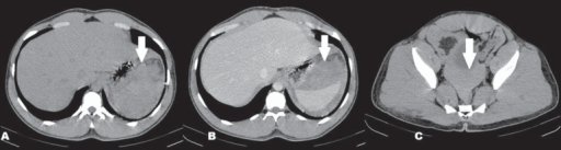
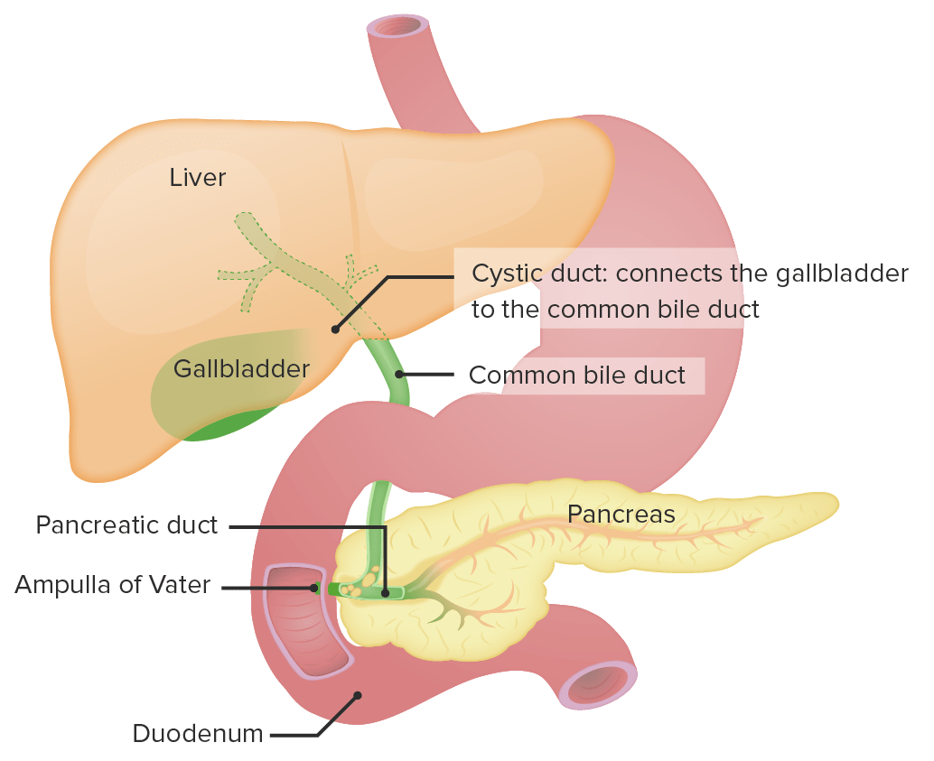
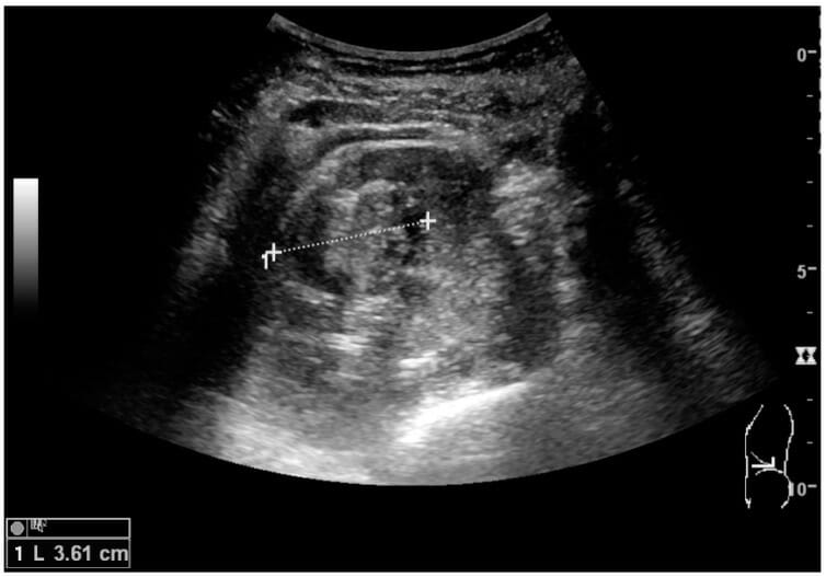
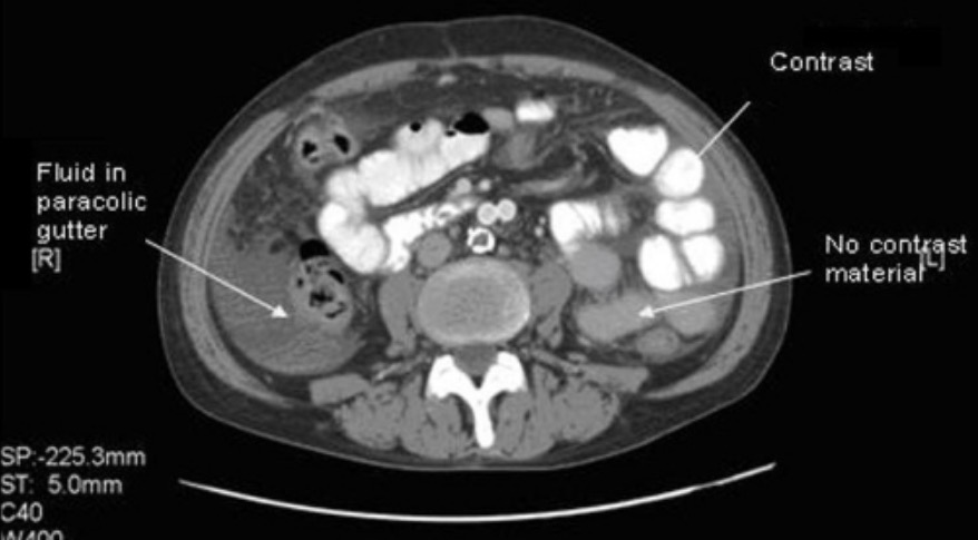
00:01 So in this lecture, we?ll be reviewing some normal abdominal and pelvic CT anatomy and we?ll be going over standard approach to when we?re looking at a CT scan. 00:11 So what I do when I look through a CT scan is I use multiple window levels and I scroll through multiple times. 00:18 It?s important to evaluate only one organ at a time. 00:21 The abdominal CT scan has a lot of different organs in there and so, if you try to take a look at everything at once, it?s very likely that you?ll miss something. 00:29 So it?s important to take a look at one organ at a time and scroll through multiple times through that organ before moving on to the next one. 00:36 By focusing on only one organ, you?ll be less likely to miss findings. 00:40 So let?s come up with the standard approach. 00:43 What I start off with is taking a look at the lung windows. 00:46 So I start off by looking at the lung bases and then I scroll all the way through the abdomen and pelvis and I look for any kind of free or abnormal collections of air. 00:54 I then look through the bone windows and I take a look for any kind of occult fracture. It?s important in all of radiology that when someone comes in with a clinical finding, you don?t focus on only that one finding. 01:05 It?s important to take a look at the entire scan and make sure that you?re seeing even the incidental findings that the patient may have. 01:11 I then take a look at the liver and I scroll multiple times through the liver. 01:15 I then go on to the spleen, the gallbladder, and the pancreas, and again, through each of these, I scroll through multiple times to make sure that I?m evaluating the entire organ before I move on to the next one. 01:27 I then take a look at the renal collecting system and the kidneys, the adrenal glands, and then I go further down and take a look at all the pelvic organs. 01:36 I then evaluate the entire mesentery, the blood vessels, and then you wanna take a look at the entire bowel. 01:43 Always check the appendix on a patient that?s coming in with abdominal pain or even on a patient that?s not coming in with abdominal pain because oftentimes, you may find an occult appendicitis that just hasn?t become symptomatic. 01:55 I then take a look at all of the soft tissues. 01:58 So lung windows are very useful in identifying small air bubbles. 02:04 So every abdominal CT should be evaluated in lung windows to look for free intraperitoneal or abnormal collections of air. 02:11 So as you can see on the single slice, this is all air that?s located within the bowel. 02:16 And let?s take a look at these CT images. 02:22 So can you see where the free air is? So here?s a collection of abnormal air. 02:33 If you compare it with the air that?s located just posterior to it, you can tell the difference. The air that?s located posterior to it appears to form within the bowel wall while the air that?s circled is actually outside of any kind of structure, and so this is how you can identify free air when you look through lung windows. 02:51 Next, I take a look at the CT scan in bone window and I evaluate all of the different bony structures. 02:58 This is just an example of a couple of bony structures that you can see. 03:01 So we have multiple ribs and then you have the vertebral body as well as the spinous process of the vertebral body. 03:10 I then go on and take a look at the liver. 03:16 So the liver is divided into right, left, and caudate lobes, and those are then subdivided into multiple segments by the vessels. 03:23 You may often hear about the quadrate lobe as well and that?s actually now known as the medial segment of the left lobe. 03:29 So the middle hepatic vein divides the liver into the right and the left lobes. 03:35 The right hepatic vein divides the right lobe into its anterior and posterior segments and the falciform ligament divides the left lobe into its medial and lateral segments. 03:46 The portal vein then divides the liver into upper and lower segments. 03:51 So this is an example of the medial segment of the left lobe of the liver. 03:56 Here, we have the right lobe of the liver. 03:59 We don?t definitely see the hepatic veins here but it?s probably located somewhere around here and you would see it as you scroll through the liver. 04:07 This is the lateral segment of the left lobe of the liver and as you remember, the falciform ligament right here is what divides the medial and lateral segments of the left lobe. 04:18 Down here, we see the caudate lobe. 04:20 So about 80% of the vascular supply to the liver is portal vein but it also has a partial supply by the hepatic artery about 20% or so. 04:31 Normally, the liver should have a very sooth contour and if it doesn?t, you wanna suspect an abnormality. 04:37 The density of the liver should equal that of the spleen. 04:40 So as you can see here, this is a normal-appearing liver which appears about the same density as a normal-appearing spleen. 04:48 The spleen is about 12cm in longitudinal dimension. 04:54 It?s actually best measured on ultrasound and it does tend to appear heterogeneous on arterial phase imaging but it should appear homogeneous on portal venous phase imaging and again, about the same density as the liver. 05:05 So here we have the pancreas. 05:09 Just to take a look at the surrounding structures, we can see here the liver which is adjacent to the pancreatic head. 05:18 Just anterior to the pancreas, we have the stomach which is partially filled with contrast here and then just posterior to the pancreas, we have the kidney here and this is the left kidney that I?m pointing at right now. 05:32 So the pancreas is not normally seen on a single slice. 05:36 You actually have to scroll through multiple slices to see the pancreas. 05:39 The normal pancreatic duct is about 3 to 4mm and in general, it shouldn?t really be visualized on a CT scan. 05:45 As you can see here, we actually don?t see a normal duct within this pancreas and if you do see it, you wanna suspect that maybe it?s slightly dilated or abnormal. 05:54 The pancreas consists of the head, which is located approximately here, the uncinate process which comes down a little inferiorly, the body, and to the tail. So each of these are general definitions. 06:10 There?s really no anatomical division here and again, it?s located in multiple planes and really can?t be seen on a single axial slice. 06:18 So now let?s move on to the gallbladder. 06:21 The gallbladder is kind of embedded underneath the liver but is pretty well seen on a CT scan. 06:27 Here we have an axial CT scan which shows you this fluid-filled sac and here we have a coronal CT scan which shows you the gallbladder as well. 06:35 It?s actually located between the right and left lobe of the liver and it?s really best evaluated by ultrasound. 06:42 So if there is an abnormality that?s suspected within the gallbladder, an abdominal ultrasound is really the first line of imaging. 06:48 The gallbladder wall should not measure more than about 3mm in thickness and again, that?s really best seen on ultrasound but maybe suspected on CT. 06:57 The adrenal glands are located above the kidneys. 07:01 They?re shaped like an upside down ?Y? and you can see them here. 07:06 They?re small and they can be sometimes hard to identify especially when there?s adjacent pathology. 07:13 So now let?s take a look at renal anatomy. 07:20 So here we have a diagram of the kidney which demonstrates a cortex right here. 07:27 We have multiple renal pyramids which comprise the renal medulla and then we have the renal hilum where we have the ureter and blood vessels going in and out of the kidney. 07:40 Here we have a portion of the proximal ureter coming out of the kidney. 07:45 So kidneys are retroperitoneal organs. 07:49 Their hilum again consists of the renal pelvis, the artery and the vein. 07:53 The pelvis is what drains out into the ureter. 07:56 Renal lesions are often localized into the upper pole, the lower pole or the interpolar region. 08:02 The interpolar region is located around the level of the hilum and again, there?s no real anatomical division but just an approximate division. 08:09 The urinary bladder is seen within the pelvis as a fluid-filled structure. 08:15 The wall is usually about 5mm or less and you can see it here in both the axial and the coronal CT scans. 08:24 So what is intraperitoneal versus retroperitoneal? The parietal peritoneum is a thin membrane that lines the abdominal and pelvic walls and the visceral peritoneum is an invagination of the parietal peritoneum that lines most of the abdominal organs. 08:41 So if a structure is located intraperitoneal, it means that the organ is lined all around by the visceral peritoneum. 08:49 If it?s retroperitoneal, that means it?s lined on only one side by the visceral peritoneum. 08:54 So there?s a long list of organs that are retroperitoneal. 08:58 I remember it by the mnemonics "SADPUCKER". 09:01 So S stands for suprarenal or adrenal glands, A is for aorta and IVC, the D is the duodenum but that excludes the first 2 to 3cm which is actually intraperitoneal. 09:13 We have the pancreas except for the pancreatic tail, the ureters, the colon predominantly the ascending and the descending colon, the transverse is actually intraperitoneal, the kidneys are retroperitoneal organs, the esophagus or the portion of the esophagus that?s within the abdomen and then the rectum. 09:33 So let?s take a look at the vessels within the abdomen. 09:37 When this scan is performed in arterial phase imaging, we can see the arteries very well and this is the coronal and a sagittal reconstruction of a CT scan performed in the arterial phase. 09:47 Here, you can see the aorta coming down, and then you can see both renal arteries. 09:52 On the sagittal view, or the view where you?re looking at the side from the side of the patient, you can again see the aorta coming down and then you have the celiac artery superiorly here and the superior mesenteric artery. As you scroll through these CT images, you?ll be able to follow each of these vessels out. 10:09 This is actually a 3D rendering that was created off of the CT scan that was performed. 10:15 In the venous phase, you can see the venous system very well. 10:21 So again, we have an axial CT image and then we have the coronal CT image in the portal venous phase and here, you can see the renal veins. 10:29 You can see a portion of the inferior vena cava and you still have a little bit of contrast within the aorta. 10:36 Here, you can actually see the inferior vena cava a little bit better on this coronal image and you can see one of the renal veins coming out. 10:44 This is again a portion of the abdominal aorta that has calcification within the walls because of atherosclerotic disease. 10:51 This is another slice within the portal venous phase and now you can see the portal vein really well adjacent to the liver hilum and then here, we have a branching into the superior mesenteric vein and the splenic vein. 11:08 So when you take a look at the bowel, you really need proper distension for accurate assessment. 11:13 The normal bowel wall thickness is about less than 3mm and as you can see here, this is a normal-appearing colon. 11:21 It actually looks like there are areas of wall thickening and some of these could be because of incomplete distension form the oral contrast and it could also be because of peristalsis of the bowel. 11:31 Here, we have an axial CT image, and you can see the difference. 11:36 So when the small bowel here is fully distended, you actually don?t see any of the walls around it. 11:42 Here, we have a section of small bowel that?s not fully distended and it appears to have wall thickening but this is probably because it?s incompletely distended. 11:50 So when you are evaluating for bowel wall thickening, you wanna look for other secondary changes. 11:54 Maybe surrounding inflammatory change would help you and you also wanna see whether that portion of the bowel is fully distended or not. 12:02 So let?s take a look at this case. 12:04 What are the different phases that are depicted here and do you see the pathology? So this is an example of renal calculi. 12:19 If you look at the first scan, this is performed without intravenous contrast and you can see the arrow points to a high-density within the kidney which represents a stone. 12:29 The second image is performed in the portal venous phase and here you can also see the stone and you can also see a little bit of contrast within the surrounding structures. 12:38 However, let?s take a look at the delayed phase. 12:40 So this is the phase that?s performed at about 10 minutes or so and you can see a lot of contrast within the renal pelvis. 12:47 This actually obscures evaluation of the stone and this is the reason why you don?t want to give contrast in a patient where you?re suspecting a renal calculus because you may actually miss the stone because the contrast will hide it. 12:58 So we?ve gone over some normal abdominal anatomy and we?ve gone over some normal techniques and a good approach to the CT of the abdomen and pelvis. 13:09 Hopefully, this will provide a good ground work as we move on to some pathology.
About the Lecture
The lecture Normal Abdominal and Pelvic CT Anatomy by Hetal Verma, MD is from the course Abdominal Radiology. It contains the following chapters:
- Lung and Bone Windows
- Liver Anatomy
- Spleen, Pancreas, Gallbladder and Kidneys
- Vessels - Arterial and Venous Phase Imaging
- Bowel
Included Quiz Questions
What segments of the liver does the falciform ligament divide?
- The medial and lateral segments of the left lobe of the liver
- The upper and lower segments of the liver
- The caudate and quadrate lobes of the liver
- The anterior and posterior segments of the right lobe of the liver
- The caudate lobe from the rest of the liver
Why are "lung windows" used in reviewing a CT scan of the abdomen?
- To check for free intraperitoneal air in the abdomen
- To evaluate the portion of the lungs that extends into the abdomen
- To check for pulmonary emboli
- To check for lung cancer metastases to the abdominal organs
- Lung windows are only used in CT of the chest, not CT of the abdomen.
What is the width of a normal pancreatic duct?
- 3–4 mm
- 2–3 cm
- 1–2 mm
- >5 mm
- 0.5–1 mm
Which organs are retroperitoneal?
- Kidneys and ureters
- Liver and gallbladder
- Head and tail of the pancreas
- Small intestines and colon
- Spleen and uterus
Why should contrast be avoided while assessing for renal calculi?
- In the delayed phase, the contrast in the renal pelvis obscures the evaluation of a stone.
- The contrast remains much longer in the kidneys due to the stone and thus can cause damage to the kidney.
- The density of the renal stone and the portal vein during the portal venous phase is the same making it difficult to assess for the stone.
- The renal stone absorbs the contrast material to a large extent and then dissolves into smaller particles causing obstruction of the kidneys.
- In the arterial phase, the stone tends to move further into the renal medulla leading to papillary ischemia.
What is TRUE regarding imaging of the liver and spleen?
- The density of the liver and spleen are equal on CT scan imaging.
- 80% of the vascular supply to the liver is from the hepatic artery and 20% is from the portal vein.
- The liver has a bumpy contour.
- The spleen is better visualized on a CT scan than ultrasound.
- The spleen is about 6 to 10 cm in size.
A newborn child with ambiguous genitalia is suspected to have congenital adrenal hyperplasia. A CT scan of the abdomen is most likely to show abnormality in which site and organ?
- An upside-down "Y-shaped" structure on the top of the kidneys
- A "bean-shaped" structure below the spleen
- An upside-down "V-shaped" structure attached to the spleen
- A "V-shaped" structure located posterior to the inferior vena cava
- A "balloon-shaped" structure attached to the liver
Author of lecture Normal Abdominal and Pelvic CT Anatomy

Hetal Verma, MD
Customer reviews
5,0 of 5 stars
| 5 Stars |
|
5 |
| 4 Stars |
|
0 |
| 3 Stars |
|
0 |
| 2 Stars |
|
0 |
| 1 Star |
|
0 |
