Playlist
Show Playlist
Hide Playlist
Abdominal CT Techniques: Hounsfield Units and Intravenous Contrast
-
Slides Abdominal CT Techniques.pdf
-
Download Lecture Overview
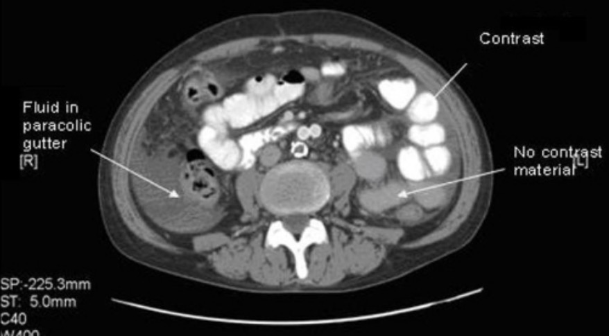
00:01 So now let's review some common CT techniques and discuss how these techniques can be manipulated to help you identify abnormalities. 00:09 Radiation is administered during a CT scan and it varies depending on the type of machine that's used, the type of scan that's being performed, and the patient's body size. 00:20 So sometimes, some of the older machines actually administer more radiation than some of the newer machines do. 00:25 Head CT scans for example will have less radiation than an abdominal CT scan just because the area being scanned is smaller in size. 00:32 Patient's body habitus can also change the amount of radiation that's administered so patient's that are larger in size need more radiation to penetrate the body size and the body tissue. 00:42 It's important to remember though that radiation is additive, so multiple scans really should be limited whenever possible. 00:49 So you really want to limit your overall lifetime dose of radiation. 00:52 So let's review Hounsfield units before we move on. 00:57 A Hounsfield unit is a measure of the density of a structure and density is the amount of radiation that that structure absorbs. 01:04 So air has the lowest Hounsfield units. It measures about -1000 Hounsfield units. 01:10 It then progresses on to fat which measures about -50 to -100, water which is about zero, soft tissue which ranges from about 20 to 300, and then bone which is the highest Hounsfield units and it measures greater than about 700. 01:26 If you put metal in there, which is not really an anatomical structure, metal will actually be the most Hounsfield units close to about a thousand. 01:33 So what are window levels? Window levels are digital manipulation of the image that help you accentuate structures of various different Hounsfield units. 01:42 Window levels can actually be changed by the radiologist as a post-processing mechanism after the CT scan is obtained. 01:48 So the CT scan is obtained only in one type of window and then everything else can be done afterwards. 01:54 So this is an example of 3 different types of window levels. 01:57 On the left we have lung windows. 01:59 It creates a very white appearance so if you're actually looking at the lungs then the lungs would be best seen on these windows. 02:05 However, these are also very useful in evaluating for free air. 02:08 The middle is the soft tissue window which is the window that's used most commonly to take a look at the solid organs of the abdomen and the right is the bony window and that's used to take a look at the bony structures. 02:19 So whenever I take a look at a CT scan I scroll back and forth using each of these windows because each window shows me something different within the abdomen. 02:28 What are some different acquisition variables? So we can use intravenous contrast, we can use oral contrast, and we can perform the CT scan at different time delays after intravenous contrast administration. 02:40 Let's take a look at when each of these would be useful. 02:43 Intravenous contrast is a low osmolar, nonionic, iodinated solution. 02:49 It actually opacifies structures based on the amount of blood flow within that structure so structures that have more blood flow will be more opacified than structures that don't have blood flow. 03:00 This is excreted by the kidneys and it can have side effects. 03:04 So it can cause acute tubular necrosis in patients that have underlying renal failure so if a patient has a GFR of less than 30 or creatinine of greater than about 1.5, intravenous contrast is contraindicated because it can cause acute tubular necrosis that may or may not be reversible. 03:21 Occasionally, it can cause hives and itching and very rarely it can cause cardiopulmonary collapse so because of these all radiology centers should be equipped with crash equipment and should be staffed by a physician who is trained in running a code. 03:36 It's actually normal for patients to have a feeling of warmth during contrast administration so when patients complain of this it really should not be confused for a contrast reaction. 03:45 Oral contrast is dilute barium sulfate and that's the one that's most commonly used. 03:52 However, gastrografin which is a water soluble type of contrast can also be used if there's suspicion of a bowel perforation and that's because if gastrografin penetrates into the peritoneum it can be absorbed and dilute barium sulfate does not. 04:05 We use approximately 1000-1500 mL of oral contrast and this is usually administered about one and half to two hours prior to CT scanning. Oral contrast is not absorbed and it does not affect the kidneys. 04:20 So if a patient has a contrast allergy, it's still safe to administer oral contrast. 04:25 So intravenous contrast is useful almost always when performing an abdominal CT. 04:30 The only exception to this is when it's being performed to detect renal calculi and we'll take a look at an example of this. 04:37 Oral contrast is useful whenever you're trying to determine any kind of bowel pathology, any kind of intraabdominal abscess, and in most instances of nontraumatic pain. 04:47 Again, the only exception is when you're trying to detect renal calculi.
About the Lecture
The lecture Abdominal CT Techniques: Hounsfield Units and Intravenous Contrast by Hetal Verma, MD is from the course Abdominal Radiology. It contains the following chapters:
- Radiation: Hounsfield Units
- Intravenous Contrast
Included Quiz Questions
What is TRUE regarding CT imaging?
- The radiation dose varies based on the type of the machine and the type of scan.
- A head CT scan has more radiation than an abdominal CT scan.
- Air has higher Hounsfield units than water.
- Fat has higher Hounsfield units than soft tissue.
- Radiation dose is the same with all machines and scans.
Which of the following is a feature of an intravenous contrast agent?
- IV contrast opacifies structures based on the amount of blood flow.
- IV contrast is ionic with high osmolarity.
- IV contrast should be avoided in patients with a GFR of <60 ml/min.
- IV contrast is metabolized by the liver and excreted in the stool.
- IV contrast is not a drug and thus does not cause allergic reactions.
Which contrast agent is used if there is a suspicion of bowel perforation?
- Gastrografin
- Dilute barium sulfate
- Gadolinium medium
- Barium iodide
- Saline water
Customer reviews
5,0 of 5 stars
| 5 Stars |
|
5 |
| 4 Stars |
|
0 |
| 3 Stars |
|
0 |
| 2 Stars |
|
0 |
| 1 Star |
|
0 |


