Playlist
Show Playlist
Hide Playlist
What is a Fracture?

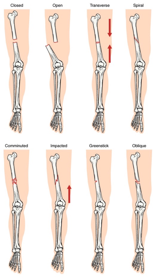
00:01 So let's move on to fractures. 00:02 What is a fracture? A fracture is actually the disruption in the cortex of a bone. 00:09 There are multiple radiographic findings to help you to help you look for fractures. 00:14 One is a linear lucency or a black line through the bone that extends out to the cortex. 00:19 Fractures usually have very sharp angles and there's no surrounding cortex at the sight of the fracture. 00:25 The fractures usually have irregular margins as well. 00:29 One thing that you wanna keep in mind, are there are other things that can actually look like a fracture. 00:35 One of them is the nutrient canal and that?s a linear lucency within the cortex of a bone that carries the blood vessels. 00:41 The difference is the nutrient canals are smooth and they have a sclerotic margin surrounding them while fractures are irregular and they don't have the sclerotic margin around them. 00:50 So let's just take a look at an example of a nutrient canal. 00:54 They're a little bit hard to see, so let's take a zoomed in look here and you can see in the green arrow pointing to a nutrient canal here. 01:01 So you can see this lucency and if you look carefully, you can actually see an area of sclerosis on both sides of this lucency. 01:09 It's very smooth and so this tells you that this is an anatomical structure. 01:14 It's just a normal nutrient canal. 01:16 Dislocation and subluxation are often seen and are often associated with fractures. 01:20 So a dislocation is when 2 bones that normally articulate with each other no longer articulate. 01:26 So they separate away. 01:28 Subluxation is when 2 bones that normally articulate with each other are now only partially in contact, so not completely dislocated but kind of halfway through. 01:37 This is an example of a dislocation at the 5th DIP joint of the foot. 01:43 So you can see that the middle phalanx here is dislocated at the DIP joint from the distal phalanx. 01:50 Normally, they should articulate the way the remainder of this DIP joints do. 01:55 There are multiple ways of describing a fracture which you wanna keep in mind is in your report when you describe a fracture, you want the person listening to your report to be able to visualize what you're seeing without actually taking a look at the pictures. 02:09 So you have to be very thorough in your description. 02:11 You wanna include the location of the fracture. 02:13 You wanna say whether it's a complete fracture or an incomplete fracture and you wanna talk about displacement. 02:19 So in terms of the location you wanna talk about which bone is involved, which part of that bone is involved and does it extend to the joint space which can have clinical implications. 02:31 So a complete fracture is one that extends all the way through the cortex. 02:36 That can also be characterized into transverse, oblique, spiral or comminuted and we'll take a look at these images. 02:44 An incomplete fracture is one that extends through only part of the cortex so not all the way through and this includes a buckle fracture or a greenstick fracture. 02:53 So incomplete fractures occur in soft bones. 02:57 They're most commonly seen in the pediatric population. 03:00 This is an example, just an overview of all the different types of fractures and their description. 03:05 And we'll go into each of this in a little bit of detail. 03:08 You can use this as a reference as we go through the lecture. 03:11 So a complete fracture includes a transverse fracture and this is when the fracture line is perpendicular to the long axis of the bone. 03:20 An oblique fracture is one in which the fracture line is diagonal with respect to the long axis of the bone and a spiral fracture is usually caused by a twisting force, so you can see it's spiraling around the long axis of the bone. 03:34 A comminuted fracture is one that produces more than 2 fragments. 03:39 So let's take a look at this x-ray of the hand. What do you see here? The abnormality is of the second digit. 03:54 So this is an example of an oblique fracture. 03:58 It's an oblique fracture of the second proximal phalanx. 04:00 You can see it right here and the fracture line is diagonal with respect to the long axis of the bone. 04:07 How about this one? So this is a radiograph of the right shoulder and this represents a comminuted fracture. 04:17 So it's a fracture that produces more than 2 fragments. 04:20 A simple fracture on the other hand is one that produces only 2 fragments. 04:24 Comminuted fractures can be further subdivided into segmental and butterfly. 04:29 So what does this look like? This is a comminuted fracture. 04:33 You can see that there are 3 discrete fragments. 04:36 So we have one, two and three. 04:39 This is an example of a segmental fracture where the central portion of the shaft becomes an isolated segment. 04:45 So incidentally, we see another finding in the tibia. 04:49 What do you think this represents? It's a very high density structure, higher in density than the surrounding bone. 05:02 So this is an example of an intramedullary rod which was placed surgically to fix the tibial fracture. 05:08 You can see a portion of the surrounding tibial fracture here. 05:11 Let's take a look at this case. 05:16 So what kind of fracture is this? This is an example of a butterfly fracture where the central portion is triangular in shape. 05:26 So this is another type of comminuted fracture with a butterfly fragment here. 05:31 So another way of describing fractures is using displacement. 05:37 So we wanna go over location. 05:39 We also wanna go over displacement and the 3 ways to describe displacement are angulation, displacement and override. 05:47 So angulation is the degree to which the distal fragment is angulated from its normal portion. 05:51 Displacement is the amount by which the distal fragment is offset from the proximal fragment either side-to-side or front-to-back and override is the amount by which the distal and proximal fragments overlap with each other. 06:06 You can also have a fracture which is impacted and that's the amount by which the distal fragment has impacted or hit into the proximal fragment and you can have distraction which is the amount by which the proximal and distal fragments are separated from each other which is the opposite of overlap essentially. 06:25 So let's take a look at this. How would you describe this fracture? So this is displacement and overriding of fracture fragments. 06:37 So we have transverse fractures of the radius and the ulna and we have full shaft radial displacement of the distal fracture fragment. 06:44 So here we have the radius and here we have the ulna. 06:50 We have transverse fractures of each one and then the distal fragment is displaced a full shaft in the radial direction. 06:58 There's approximately 1 centimeter of overriding of fracture fragments as well. 07:04 Let's take a look at this case. 07:12 So we have a radiograph of the right hip and the arrows point to 2 abnormalities that are seen here. This is an example of an avulsion fracture. 07:22 There's an avulsion fracture of anterior superior iliac spine which is the insertion site of the sartorius muscle and then there's also an avulsion fracture at the level of the lesser trochanter which is the insertion site of the iliopsoas muscle. 07:40 An avulsion fracture is when a small piece of bone is pulled off the parent bone by a ligament or a tendon and that's what happens in this case. 07:48 One thing that you wanna keep in mind when you take a look at an avulsion fracture, again there are anatomical structures that can look somewhat similar. 07:56 So there are accessory ossicles. 07:58 Those are bone fragments that form from a secondary ossification center and they don't fuse with the parent bone. 08:03 So they remain a separate ossified fragment and then you have sesamoids that are small bones that form within a tendon. 08:11 Both of these can look very similar to an avulsion fracture. 08:14 The difference is that accessory ossicles and sesamoids are smoothly marginated and they contain a cortex all the way around while fracture fragments from an avulsion fracture are irregular and they will not have a cortex all the way around. 08:26 This is an example of an accessory ossicle. 08:30 It's a well corticated os trigonum and this is a common location for an accessory ossicle. 08:37 They can be found in a variety of different locations within the body.
About the Lecture
The lecture What is a Fracture? by Hetal Verma, MD is from the course Musculoskeletal Radiology. It contains the following chapters:
- Basics of Fractures
- Complete vs. Incomplete Fracture
- Displacement
Included Quiz Questions
What structure can appear similar to a fracture on an X-ray?
- Nutrient canals in the cortex
- Epiphysis of the femur
- Multiple myeloma of the skull
- Ewing sarcoma of the right femur
- Anterior dislocation of the shoulder
Which statement is FALSE regarding fracture?
- A buckle fracture is a complete fracture extending through the entire cortex.
- A greenstick fracture extends through a part of the cortex.
- It is important to describe the location, displacement and the type of fracture.
- A complete fracture extends through the entire cortex.
- A spiral fracture is caused by a twisting force.
Segmental and butterfly fractures are two types of…?
- …comminuted fractures.
- …transverse fractures.
- …oblique fractures.
- …spiral fractures.
- …incomplete fractures.
The amount by which the distal and proximal fragments overlap with each other is known as…?
- …override.
- …angulation.
- …distortion.
- …displacement.
- …effacement.
Author of lecture What is a Fracture?

Hetal Verma, MD
Customer reviews
5,0 of 5 stars
| 5 Stars |
|
1 |
| 4 Stars |
|
0 |
| 3 Stars |
|
0 |
| 2 Stars |
|
0 |
| 1 Star |
|
0 |
everything is well explained and organised. it isn't overcomplicated and is very well taught
