Playlist
Show Playlist
Hide Playlist
Eyeball and Retina
-
Slides 15 VisualPathway BrainAndNervousSystem.pdf
-
Download Lecture Overview
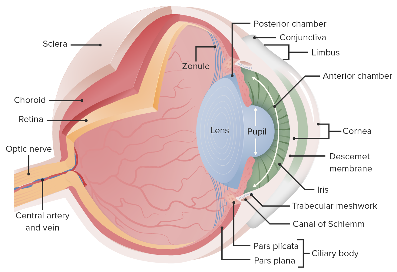
00:01 Welcome to this presentation on the visual pathway. 00:08 The first topic that I'm going to guide you through on this journey along the pathway is the passage of light through the various components of the eyeball. 00:21 And this is necessary to focus the light on the phobia centralis the point at which we have the greatest visual acuity. 00:29 The first structure that light will pass through is that of the cornea, which you see labeled here. 00:38 Next, the light will pass through the aqueous humor. 00:43 And then through an aperture of the iris that we call the pupil. 00:48 And you see that opening labeled here. 00:51 The lens is the next structure through which light will pass along it's travel. 00:56 And then its greatest journey is going to be through the vitreous humor. 01:01 And then the light will hit the retina. 01:05 And by striking the retina photoreceptor cells will become activated. 01:13 At this slide depicts the major layers of the retina that will allow the visual scenes to be delivered through the optic nerve. 01:25 First, the pigment epithelium is shown here. 01:30 This is the external most component. 01:34 The purpose of the pigment epithelium is to absorb any scattered light, which helps with visual acuity. 01:42 The photoreceptors are shown here. 01:45 And we have cones and rods in through here. 01:49 And we'll speak to those in detail shortly. 01:54 From here, photoreceptors will activate stimulate bipolar cells. 02:03 And then the bipolar cells will communicate and activate ganglion cells. 02:10 And then the axons from these ganglion cells will then form the optic nerve. 02:16 And the optic nerve is forming right here on the right upper portion of this image. 02:23 Now, let's take a look in greater detail about the photoreceptors. 02:30 First, we have the rods. 02:33 And here's a nice illustration demonstrating the rod. 02:42 And then the cones are shown here at least one cone And you can see this cone shaped appearance here to the outer segment of this photo receptor. 02:56 Now, let's describe the functions of the cones and the rods. 03:04 The cones are located specifically within the fovea centralis exclusively, there are no rods at this place. 03:12 As you move outside of the fovea centralis you're within the macula lutea. 03:17 The cone density will start to decrease and then rods will start to increase in density. 03:23 And then once you get to the periphery of the macula lutea then you have exclusively rods in the peripheral aspects of the retina. 03:35 Cones are necessary to operate in bright light. 03:41 They are responsible for conferring color vision. 03:45 And in order to do so there's a population of red, green, and blue cones. 03:51 They are responsible for high visual acuity. 03:56 And if we have an excessive loss of cones, this is a cause of legal blindness. 04:07 Rods which start to show up outside of the fovea centralis and then we see them exclusively outside of the macula are responsible for low light vision. 04:23 As a result, they have poor visual acuity and they are responsible, for a chromatic vision. 04:31 So we see scales or shades of gray and not color. 04:36 And excessive loss of rods impairs our ability to see in low light or during nighttime does this can be a cause of night blindness. 04:52 The retina in certain situations can become detached And when that happens there is a separation of the neuro retina, which would be the boundary between the photoreceptors and the pigment epithelium. 05:14 As a result to this separation, the neural retina from the pigment epithelium blood supply to the photoreceptor cells is disrupted. 05:23 The blood supply to this area is from blood vessels that reside in the choroid which will be down in this area here. 05:32 And so you are dependent then on the diffusion of the components from the blood to maintain the viability of the retina that becomes lost when we have detachment. 05:45 Possible symptoms are a shower of pepper or floaters as visual sensations. 05:51 This is do extravasated red blood cells that came from a disruption of the blood supply. 05:56 You may also see flashing lights, or a shadow, or curtain over the field of vision. 06:02 The retinal detachment can result from different causes. 06:06 The most common one is rhegmatogenous retinal detachment, which is a structural collapse of the vitreous humor that pulls the retina away from the retinal pigmented epithelium. 06:17 Then there's a tractional retinal detachment where scar tissue exerts tractional force and is pulling the retina away from the retinal pigmented epithelium. 06:28 This may be seen in diabetes for example. 06:31 Another cause is exudative retinal detachment. 06:35 In this case, fluid leaks from blood vessels and separates the retina. 06:40 It may be caused by inflammation or abnormally leaky blood vessels.
About the Lecture
The lecture Eyeball and Retina by Craig Canby, PhD is from the course Visual Pathways. It contains the following chapters:
- Eyeball
- Retina
- Retina – Cones
- Retina – Rods
- Retinal Detachment
Included Quiz Questions
When certain cells are lost in excessive quantities, night blindness may occur. Which of the following cells would cause this condition?
- Rods
- Cones
- Ganglion cells
- Bipolar cells
- Pigment epithelium
Which of the following structures contains only cones?
- Fovea centralis
- Bipolar cell layer
- Pigment epithelium
- Macula densa
- Macula lutea
Which of the following cells that are extravasated cause the sensation of “floaters” in patients with retinal detachment?
- RBCs
- Rods and cones
- Macrophages
- Lymphocytes
- Neutrophils
Which of the following cells do photoreceptors stimulate in the retina?
- Bipolar cells
- Rods and cones
- Optic nerve cells
- Pigment epithelium cells
- Ganglion cells
Customer reviews
4,0 of 5 stars
| 5 Stars |
|
1 |
| 4 Stars |
|
0 |
| 3 Stars |
|
1 |
| 2 Stars |
|
0 |
| 1 Star |
|
0 |
This is nice a clear and well paced lecture. It's easy to understand. Thanks.
I think you could have gone in a little more in depth into the structure of photoreceptors. There were also a few other cells, such as the horizontal cells which backpack onto parts of the visual pathway-this knowledge is essential to understanding concepts of contrast. overall i do think it was a good lecture, but a little more depth into the vitreous humour and the aquous humour would have also helped


