Playlist
Show Playlist
Hide Playlist
Spinal Trauma

-
Slides Spinal Trauma.pdf
-
Download Lecture Overview
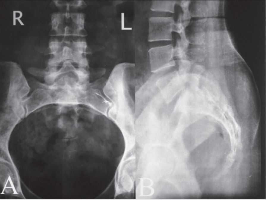
00:01 So in this lecture we'll be discussing spinal trauma. 00:04 When a patient comes in with a traumatic injury to the spine, imaging can play a key role because it can help you exclude an abnormality that can lead to a long-term complication in the patients such as paralysis. 00:15 So when a patient comes in with a cervical spine trauma if there's any kind of suspicion of a spinal fracture, the patient's neck should not be moved because an unstable fracture can compromise spinal cord integrity. 00:28 So the first thing that's done is a trauma protocol which involves the cross table lateral radiograph of the neck and this ensures that there's no fracture or subluxation of the cervical spine prior to obtaining further imaging which may involve manipulation of the patient's neck. 00:43 Common mechanisms of spine trauma include a flexion injury, an extension injury, a rotational injury or a shearing abnormality. 00:53 So these are some common spinal fractures. 00:56 The most common fracture of the spine is the compression fracture. 00:59 We can also have what's called a burst fracture. 01:02 A burst fracture is caused by an axial loading injury in which the disk above is pushed into the vertebral body below. 01:09 This can result in a comminuted fracture of the vertebral body and it's often associated with retropulsion of bony fragment into the spinal canal which results in an unstable fracture that can cause neurologic deficits. 01:20 So this is an example of a radiograph and the CT scan that demonstrates the burst fracture. 01:27 So you can see that there's a comminuted compression fracture of the vertebral body and there?s displacement of bony fragments posteriolary into the spinal canal. 01:37 So if you look at the posterior vertebral line here, when you get to the level of the fracture it's displaced posteriorly indicating posterior retropulsion of fragments. 01:46 On the CT scan you can see that there's a fracture of the vertebral body and the fragment is pushed posteriorly into the spinal canal. 01:54 There's what's called a chance fracture. 01:57 And a chance fracture is also called a "seatbelt fracture," and it's usually caused by a lap belt in a motor vehicle accident. 02:04 This is a type of flexion distraction injury and you have fracture through the vertebral body, the pedicles and the spinous process. 02:12 This can actually be associated with an increased interspinous distance and you have separation of the facet joints. 02:18 So this is also very unstable fracture and it can be associated with neurological deficits. 02:23 Often this is associated with abdominal visceral injuries as well because of the lap belt. 02:28 So here we have two CT scans, sagittal images which demonstrates a fracture through the vertebral body and into the spinous process. 02:38 So this is an example of a chance fracture often these are associated with an epidural hematoma and possibly a cord contusion as well. 02:46 So Jefferson fractures are axial loading injuries. 02:50 You have offset of the lateral masses of C1 with respect to the lateral margins of C2 when you do an open mouth radiograph of the cervical spine. 03:00 There also fractures of the anterior and posterior arches of the C1 ring and this is best seen on a CT scan. 03:06 Usually this are not associated with the neurological deficits. 03:09 So let?s take a look at this example of a Jefferson fracture. 03:13 This is what's called an open mouth view and it demonstrates lateral displacement of the lateral masses in the Jefferson fracture. 03:21 So here we have lateral displacement of the two masses laterally. 03:24 When you compare it with the lateral margins of the C2 vertebral body, you can see the prominence of the lateral displacement here. 03:32 When you compared with a normal film here, you can see that this is well align. So the lateral margins of C1 and the lateral margin of C2 is actually well aligned. While with the Jefferson fracture it's not and you see a step off on both sides. 03:46 On a CT you can see a fracture involving the anterior and posterior arch of C1. 03:52 So here we have a fracture involving the anterior arch and then here we have a fracture involving the posterior arch. 03:59 So when you're taking a look at a CT and you see a fracture of either the anterior or posterior arch, you wanna take a look for a second fracture because this is a ring, usually rings fracture in two different places not just one. 04:11 So if you see one you may be missing a subtle second fracture. 04:14 A Hangman's fracture is actually a hyperextension compression injury. 04:19 So this can occur in patients such as an unrestrained passenger who hits their forehead on a windshield in a motor vehicle accident. 04:26 This is not truly associated with a judicial hanging, which actually ends up causing hyperextension resulting in distraction of C2 and C3 and distraction of the cord. Imaging findings include a fracture of the posterior elements of C2 and the posterior aspect of C2 is actually separated from the anterior aspect of C2. 04:48 So this results in anterior displacement of C2 on C3. 04:52 This is actually not usually associated with neurological deficits. 04:55 So this is an example of a hangman's fracture. 04:58 This is a lateral cervical radiograph which demonstrates posterior displacement of the spinolaminar line. So as you can see here the line drawn with a posterior displacement right here and then again it moves anterior here. 05:11 The line should actually remain symmetric all the way up and down. 05:14 You also have anterior displacement of the C2 vertebral body and you have fracture of the posterior elements of C2. 05:22 So we've gone over several of the common spinal fractures that you may encounter. 05:27 Again, these are very important to recognize because they may have a chronic neurological deficits associated with them.
About the Lecture
The lecture Spinal Trauma by Hetal Verma, MD is from the course Neuroradiology.
Included Quiz Questions
Which of the following applies to a Jefferson fracture?
- Imaging demonstrates displacement of the lateral masses of C1 with respect to the lateral margins of C2.
- It involves the spinous process of C2
- It is caused by a flexion-distraction injury
- It is caused by a hyperextension compression injury
- It is an unstable fracture associated with neurological deficits.
A young man was an unrestrained passenger and hit his forehead to the windshield when his car was hit head-on. Imaging findings show a fracture of the posterior elements of the C2 vertebra due to this hyperextension injury. What is this fracture called?
- Hangman’s fracture
- Burst fracture
- Chance fracture
- Jefferson fracture
- Jones fracture
What is TRUE regarding spinal trauma?
- Trauma protocol involves a cross-table lateral X-ray of the neck to ensure there is no fracture or subluxation.
- A compression fracture results in a comminuted fracture of the vertebral body.
- A burst fracture is due to hyperextension injury.
- A Chance fracture is due to axial loading.
- Hangman’s fracture is the most common spine fracture associated with neurological deficits.
Author of lecture Spinal Trauma

Hetal Verma, MD
Customer reviews
5,0 of 5 stars
| 5 Stars |
|
1 |
| 4 Stars |
|
0 |
| 3 Stars |
|
0 |
| 2 Stars |
|
0 |
| 1 Star |
|
0 |
El tema fue dado de una forma concisa y clara, permitiendo comprender las diferencias entre las lesiones habladas en el video.
