Playlist
Show Playlist
Hide Playlist
Hand – Bones and Surface Anatomy of Upper Limb

-
Slides 01 UpperLimbAnatomy Pickering.pdf
-
Download Lecture Overview
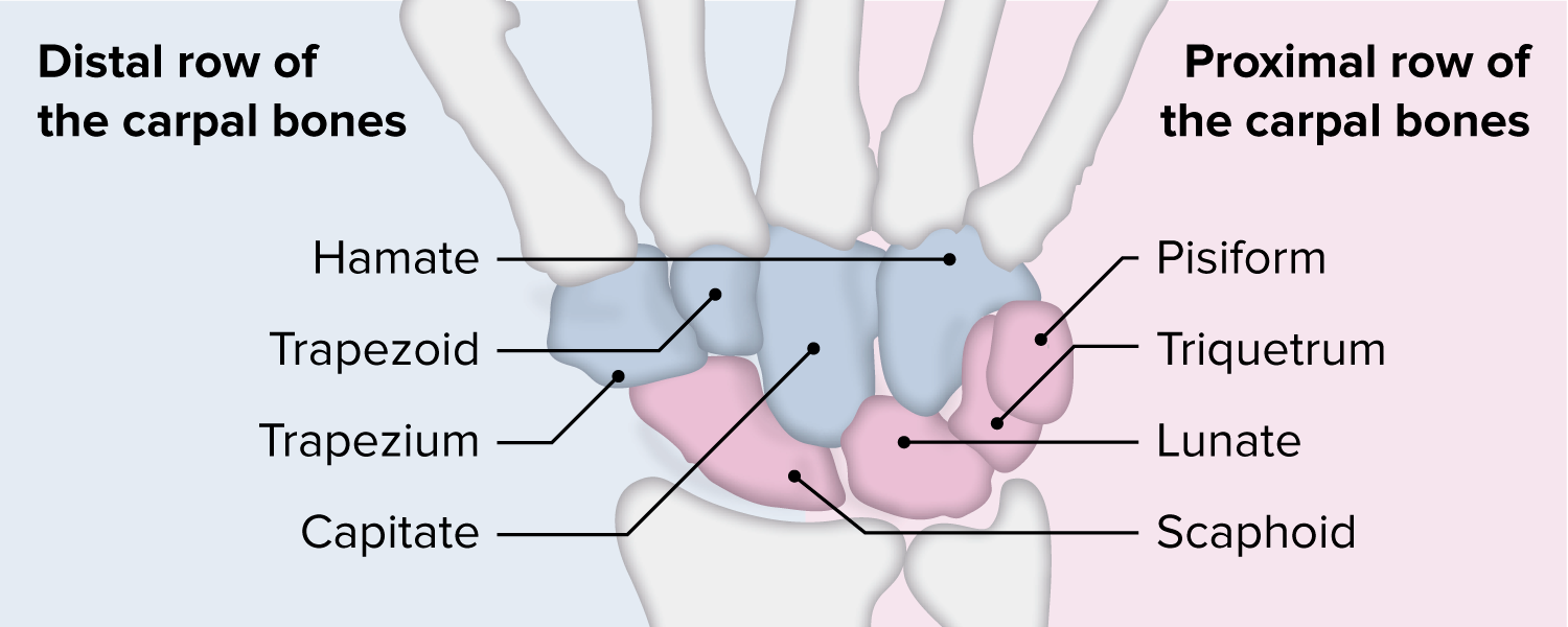
00:00 Okay. We now move into the hand. Then we can see we have a whole series of bones which are forming the wrist joint here. These are known as carpal bones. We can see on the picture, we then have five metacarpals we can see running down in this direction. And then we can see we have phalanges that give rise to digits. So let’s just look at the carpals first of all. And there are eight carpal bones and these eight carpal bones form two rows, with each rows having four carpal bones in them. And this arrangement, this high number of bones allows greater flexibility. 00:43 So there is a high level of flexibility within the wrist joint. Again allowing great movement of the wrist to assume numerous positions. We have the proximal row and from lateral to medial. So here we've got the anterior view of the right hand. So this is going to be lateral. 01:08 We can see here the first digit, the thumb, this is lateral and this fifth digit is medial here. And this proximal row from lateral to medial, we have these four bones. We have the scaphoid, we have the lunate, we have the triquetrum and we have the pisiform bone. And we can see these here. We can see we have got the scaphoid, we can see we have the lunate, we can see we have the triquetrum, and then we can see we have the pisiform. So 1,2,3,4. These four bones that form this proximal row. We can see this on this anterior view and we can also see them on this posterior view. Again remember the thumb here is lateral, so from lateral to medial, we have scaphoid, we have lunate, we have triquetrum and we have pisiform. These four bones that make up the proximal row. We also have a distal row and these are formed by trapezium, trapezoid, capitate and hamate. And these are the distal rows and again we can see them from lateral to medial, we can see the trapezium, we can see the trapezoid, we can see the capitate and then we can see the hamate with the hook of the hamate as a prominent feature. 02:35 We can see this on the anterior aspect, trapezium, trapezoid, we can see the capitate and then we can see the hamate bone. So we've got this proximal row and then we've got this distal row. We have these two rows with four bones forming each row, while we can see them both from the anterior and the posterior view. Here in the posterior view, we can see trapezium, trapezoid, capitate and hamate. 03:07 And these bones, these 8 carpal bones offer great flexibility. 03:11 If we look at the palm of the hand, this is known as the metacarpus, it is formed by five metacarpals, and these metacarpals all have a similar feature. The five of them, they have a base. We can see a base here, here, here, here and here. And the base of the metacarpals articulate with the distal row of the carpal bones. So these five metacarpals, their bases articulates with the trapezium, the trapezoid, the capitate and the hamate with the proximal row of carpal bones articulating with the radius at the wrist joint. We have a nice shaft here which we can see and then we have a head. And the head of the metacarpals form the knuckles which you can see on your own hand and these articulate with the proximal phalanges of the digits. So, each individual metacarpal will have a base, a shaft and a head. 04:15 The head articulates with the proximal phalanges. 04:19 And let's look at these phalanges. For digits 2, 3, 4 and 5, there are 3 phalanges. So digits 2, 3, 4 and 5, we have 3 phalanges. We have a proximal, we have a middle and we have a distal. And each one of these phalanges again just like a metacarpal is going to have a base, is going to have a shaft and is going to have a head. Now digits 2, 3, 4, and 5 have 3 phalanges. Digit 1 only has two. It just has a proximal and a distal. 05:02 So there we have the various bony features that make up the hand. The carpals, the metacarpals and the phalanges.
About the Lecture
The lecture Hand – Bones and Surface Anatomy of Upper Limb by James Pickering, PhD is from the course Upper Limb Anatomy [Archive].
Included Quiz Questions
How many carpal bones are there?
- 8
- 10
- 9
- 7
- 6
What is the correct arrangement of the carpal bones in the proximal row going from the lateral to the medial direction?
- Scaphoid, lunate, triquetrum, and pisiform
- Lunate, scaphoid, triquetrum, and pisiform
- Scaphoid, triquetrum, lunate, and pisiform
- Pisiform, triquetrum, lunate, and scaphoid
- Triquetrum, pisiform, scaphoid, and lunate
Which of the following bones has a prominent hook?
- Hamate
- Scaphoid
- Lunate
- Trapezium
- Capitate
Author of lecture Hand – Bones and Surface Anatomy of Upper Limb

James Pickering, PhD
Customer reviews
5,0 of 5 stars
| 5 Stars |
|
2 |
| 4 Stars |
|
0 |
| 3 Stars |
|
0 |
| 2 Stars |
|
0 |
| 1 Star |
|
0 |
good lecture, like the breakdown of structures and in relation to other ones.
1 customer review without text
1 user review without text
