Playlist
Show Playlist
Hide Playlist
Eosinophilic Lung Disease (ELD)

-
Slides ExtrinsicLungDiseases RespiratoryPathology.pdf
-
Download Lecture Overview
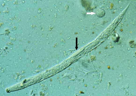
00:00 Lets take a look at Eosinophilic lung disease now. And before we move on though, the segway here, is the fact that it’s, well, for the most part, once again, hypersensitivity in exposure. However, with hypersensitivity pneumonitis, you did not necessarily see eosinophils. Is that understood? If that is not clear and you don’t have a firm grasp of that, go back and take a look at that table, so that you’re… that and the criteria. That would be the two most important. 00:31 Now, under Eosinophilic lung disease, this means that the patient was exposed to, we’ll take a look at the number of issues in which now the patient has developed what? Eosinophilia. 00:42 Clear? Defining characteristics needed to diagnose Eosinophilic lung disease (ELD) include, number 1, peripheral blood with eosinophilia. So, you have an increase in WBC, and specifically it will be increase in eosinophils. With abnormalities of pulmonary imagining. Who are you going to… what part of lung are you going to affect more so? Good. The interstitium. 01:05 The lung tissue eosinophilia demonstrated in transbronchial or open lung biopsies. 01:11 Eosinophil is the name of the game here, and increased eosinophils in bronchioalveolar lavage. Stop here. Would you tell me. Lets say that your patient was growing birds or raising birds and your patient maybe was working in a type of a silo, whatever it may be. That’s hypersensitivity pneumonitis. They’re not feeling too well. 01:35 On bronchoalveolar lavage, what you’re going to find there? Good. A low CD4 to CD8 count. 01:41 That was criteria number two. Here, with the etiologies and differentials that I’ll give you for Eosinophilic lung disease. Here, the bronchioalveolar lavage will show you, eosinophils. 01:53 All three bullet points: blood, biopsy, lavage – esosinophils. 02:02 What are my differentials? Lets begin. Drug-induced, toxin-induced. NSAIDs. You’ve heard of patients who is “allergic to drugs” such as NSAIDs. The usual suspects here for antibiotics, especially sulfonamides, penicillins, phenytoin, maybe even L-tryptophan. Good. 02:23 These will be one’s in which, upon exposure, the patient has now developed Eosinophilic lung disease. Next. 02:32 So those were drugs. What if your patient... let’s walk through the various types of, what’s known as Loeffler’s syndrome. So these will be helminths. For example, Ascaris lumbricoides. Crazy worm. Where is it? In my intestine. How big is it? I don’t know, it’s huge. What does it do? It’s like an alien in your intestine and it burrows through the intestine. Gets into where? The blood. And it goes where? Transpulmonary. Would you tell me as to, well, what you require to properly kill your helminth? Oh yeah, you need eosinophils Dr. Raj. There you go. 03:17 So, here we have Eosinophilic lung disease which is caused by what? Helminthic infections. 03:24 Understand something. If it doesn’t invade… what do you mean doesn’t invade? Where did you find this Ascaris initially? In the intestine. If it doesn’t invade, you are not going to find eosinophilia. Is that understood? An important clinical distinction. 03:39 If this thing actually invades through the intestine. Here it comes, you see it? Here I come, I’m coming into the blood and I’m going to the lung, then you’ll have eosinophilia. That must be understood in many of these helminths. Lets continue. 03:51 Hookworm. We have Ancyclostoma duodenale. We have Strongyloides stercoralis. What does this one do? This thing is so strong that it will actually penetrate through your foot when you step on it. So therefore this might be a patient who has HIV, and immunocompromised. 04:10 Imagine this thing burrowing through your foot, the sole of it, my goodness, your sole. 04:16 Not this one, but the sole, underneath your foot is so thick and this strong worm can get through? Yeah, it can. Gets into blood, transpulmonary. What’s this called? Loeffler syndrome. Let me give you a little bit more information that you will find to be interesting. Lets continue. 04:33 Tropical. Well here, you end up having what? Wuchereria bancrofti. What does this organism do? It literally blocks off, whom? It blocks off the lymph nodes, doesn’t it? So, if you block off your lymph, then what then happens? You have lymphedema. Sometimes this lymphedema is so bad that it’s amazing actually that, you can find patients in developing countries in which, I’m gonna be a little exaggerated here, but they’re literally, they are putting their scrotum in a wheelbarrow and bringing it in into the clinic. That is rather large. Welcome to elephantiasis is what I’m getting at. Could I be any more dramatic? So, welcome to Loeffler. Look for transpulmonary. 05:17 Another one simple, Brugia malayi. 05:20 Another one will be, this is a good one. A direct lung parenchymal invasion and this is called paragonimiasis. This is directly lung parenchymal invasion whereas the other ones result in eosinophilia via transpulmonary. Is that clear? Then we have the ones that love to go through haematogenous. So heavy haematogenous seeding helminth include your Trichonella, the disseminated strongyloid, the visceral larva migrans, cutaneous and schistosomiasis. So these are important ones from microbiology that you’ve covered. 05:55 I would know some of these and as to how they’re involved with different manifestations in your patient and, ultimately, in the lung, may result in what kind of disease? Eosinophilic lung disease. Often times, you call this Loeffler. 06:09 Now, there is an important point that I need to bring to your attention. If you have such strict, intense eosinophilic reaction taking place in the lung known as Loeffler, is it possible that you might then also affect your adjacent structure? What is then adjacent to the left lung? Right here, by the apex, fifth intercostal space. The heart. 06:34 And so therefore, what if you had Eosinophilic heart disease? You know what that’s called? You do. That’s called Endomyocardial fibrosis. Right? In the US, extremely uncommon, but if you take some of these tropical countries, interesting, huh? Tropical countries, which is kind of like what we see here with these helminths. Then could you have eosinophilic damage to the heart? Yes, you could. What’s that called again? That’s called Endomyocardial fibrosis. Now, be careful, because at some point, you need to distinguish that from what’s called your Endocardial fibroelastosis and that’s found with your young infants. Lets continue. 07:19 Now, here we have what’s known as your acute eosinophililc type of pneumonia. And the acute eosinophilic type of pneumonia, as you can expect it to be is rather rapid. Patients have been fire-fighters during 9/11, HIV-patients and smokers. So during this time, lets say that… crazy, a building just collapsed during 9/11, you’re a fire-fighter in the middle of that, trying to save. What are you going to breathe in? All kinds of things. 07:49 And so during this time when you’re breathing in who-knows-what, you might then have a acute eosinophilic type of reaction. Is that understood? Rapid development of acute respiratory failure. 08:03 HIV-patient, immunocompromised, smokers, perhaps. They will have acute febrile illness for less than a week’s duration. And once again here, it will be non-productive cough because it’s the interstitium that is being involved. Acute eosinophilia. Acute type of conditions. 08:21 Emergency type of fire-fighters or emergency type of calls that are taking place. 08:28 The chronic, as the name applies. 50-60% of your patients will then exhibit some type of atopic reaction. What does this mean? This would mean this individual was exposed to some type of allergen. What does atopic mean? Remember atopic asthma, in obstructive? Isn’t that the majority of your asthma patients? Of course, it is. With atopia or, excuse me, atopy, then that patient may also have atopic type of eczema. Asthma, eczema. Long period of time, been exposed to allergen, now developing what? Eosinophilic. Now, be very careful because if this patient was, once again, working as a farmer, that’s a hypersensitivity pneumonitis. 09:14 You see that? That is not Eosinophilic lung disease. You must keep that separate. 09:17 Next, with chronic type of eosinophilic, predominantly occurs in women, non-smokers and some of them been exposed to post-radiation breast cancer therapy. Huge. Radiation therapy for breast. 09:32 Chronic eosinophilic lung disease. 09:36 Next. Well, under this, now we have a condition in which we have huge granulomas. So this then brings us to Eosinophilic granulomatosis with polyangiitis. In other words, this is Churg-strauss. Now with Churg-strauss, a couple of things that I wish to bring to your attention. First and foremost, it is P-ANCA +. What does P-ANCA stand for? What’s that P stand for? It means perinuclear and it stains for MPO which is myeloperoxidase. 10:03 Is that clear? So, another name for P-ANCA, as you know, is MPO, myeloperoxidase. Whenever you think about ANCA +, you should be thinking about your blood vessels that are affected and also other organs. No exception. Vasculary disorder characterised by, sinusitis, asthma, peripheral blood eosinophilia. Do you see which one of these condition, oh, excuse me, which one of these presentations is not found in Granulomatosis with polyangiitis, formally known as Wegener? Obviously, the eosinophilia, right? Lets continue. 10:40 Here’s the sole form of vasculitis that is associated with eosinophilia and lung involvement. 10:45 The sole type of lung disease. The integumentary, cardiovascular system, GI, renal, neurologic systems may also be involved. Remember, these ANCA + can affect many organs. 10:58 Even in Wegener, now currently known as Granulomatosis with polyangiitis, which is a C-ANCA +, another name for C-ANCA, proteinase 3. Do you understand with every little thing that we do? It’s about me making sure that I tie it in to other differentials and other things that you see around in medicine, but you have to have a proper way of organising things. Let me be your anchor so that I can get you where you need to be. Lets continue. 11:26 Now, you have Allergic bronchopulmonary aspergillosis (ABPA). What is this patient exposed to? Good. 11:34 Aspergillus, fumagatus perhaps. What does it do? It then elicits a immunologic eosinophilic type of reaction. Now this is usually in a backdrop of a patient who already kinda has an atopic type of condition. So this might be asthma, or patient that might have cystic fibrosis. That’s an important one where it colonises the airway, you’re not able to properly get rid of your organism. It’s stuck. Obstructive, asthma, cystic fibrosis. I don’t have my mucociliary clearance. The histologic findings of, pay attention, bronchocentric granulomatosis is important. Bronchiolitis and eosinophilic pneumonia and mucoid impaction can lead to bronchiectasis. What does that mean to you? Infection, foul-smelling sputum, fibrosis and respiratory compromise. Be careful. Go through that order, bronchocentric type of granulomatosis, you end up destroying your bronchioles, yet once again. Bronchiolitis eosinophilic pneumonia. This is allergic bronchopulmonary aspergillosis.
About the Lecture
The lecture Eosinophilic Lung Disease (ELD) by Carlo Raj, MD is from the course Disorders of the Pulmonary Circulation and the Respiratory Regulation.
Included Quiz Questions
Which of the following are the defining criteria to diagnose eosinophilic lung disease?
- Peripheral blood eosinophilia, transbronchial or open lung biopsy eosinophilia, and bronchoalveolar lavage eosinophilia
- Peripheral blood eosinophilia, transbronchial or open lung biopsy eosinophilia, and bronchoalveolar lavage neutrophilia
- Peripheral blood eosinophilia, transbronchial or open lung biopsy eosinophilia, and bronchoalveolar lavage lymphocytosis
- Peripheral blood lymphocytosis, transbronchial or open lung biopsy eosinophilia, and bronchoalveolar lavage eosinophilia
- Peripheral blood eosinophilia, transbronchial or open lung biopsy lymphopenia, and bronchoalveolar lavage eosinophilia
Which of the following is least likely to cause eosinophilic lung disease?
- D-tryptophan
- Nitrofurantoin
- Phenytoin
- L-tryptophan
- NSAIDs
What is the name of the heart disease that is caused due to eosinophilic lung disease?
- Endomyocardial fibrosis
- Endocardial fibroelastosis
- Endocardial fibrosis
- Myocardial fibrosis
- Pericardial fibrosis
Which of the following infections causes eosinophilic lung disease by the direct invasion of lung parenchyma?
- Paragonimiasis
- Trichinellosis
- Wuchereria bancrofti
- Schistosomiasis
- Strongyloidiasis
Which of the following organisms is least likely to cause transpulmonary passage of larvae?
- Schistosomiasis
- Strongyloides stercoralis
- Ancylostoma duodenale
- Ascaris lumbricoides
- Necator americanus
Which of the following is not true regarding eosinophilic lung disease?
- Low CD4/CD8 ratio
- Eosinophilia in peripheral blood smear
- Eosinophilia in lung biopsy
- BAL eosinophilia
- History of helminth infection
Which of the following is not a clinical characteristic of acute eosinophilic pneumonia?
- > 7 days duration
- < 7 days duration
- Shortness of breath
- Dry cough
- History of HIV and smoking
Which antibody is found in eosinophilic granulomatosis with polyangiitis?
- Perinuclear antinuclear cytoplasmic antibody
- Cytoplasmic antinuclear cytoplasmic antibody
- Anti-proteinase 3 antibody
- SSB antibody
- Anti-dsDNA antibody
Which of the following is not associated with allergic bronchopulmonary aspergillosis?
- Caseating granulomatosis
- Aspergillus fumigatus
- Bronchocentric granulomatosis
- History of cystic fibrosis
- History of asthma
Author of lecture Eosinophilic Lung Disease (ELD)

Carlo Raj, MD
Customer reviews
5,0 of 5 stars
| 5 Stars |
|
5 |
| 4 Stars |
|
0 |
| 3 Stars |
|
0 |
| 2 Stars |
|
0 |
| 1 Star |
|
0 |
