Playlist
Show Playlist
Hide Playlist
Testis: Seminiferous Tubule – Male Reproductive System

-
Slides 06 Human Organ Systems Meyer.pdf
-
Download Lecture Overview
00:01 important to point out their associated with the testis at this stage. Let’s look at this seminiferous epithelium. It’s the epithelium, as I pointed out, that gives rise to spermatozoa. 00:11 On the left-hand side is a section taken through the testis. You can see a number of profiles taken through these tubules. And within the tubules, you can see the epithelial cells, the spermatogenic cells. You can also see spaces between the tubules. Some of that space, you can see connective tissue, which will be part of the connective tissue septa that creates the lobules. 00:39 Other parts will be what we call interstitial space. And that interstitial space is very important that I’ll point out later on because that interstitial space houses steroid-secreting cells called Leydig cells. Turn your attention now to the right hand of the slide and look at the diagram. That diagram is going to persist in the next few slides when I describe the process of spermatogenesis. It first of all, has on the very base of the diagram a fibroblast and a basal lamina that represents part of the wall of each seminiferous tubule. 01:24 And then as you move towards the top of the slide, it shows you the different processes that the spermatocytes go through from being a spermatogonia, the germ cell, until they’re finally, at the top, released as spermatozoa. And the different colors there relate to various stages of their differentiation. They actually relate to the various stages of the meiotic process as well that I’ll point out. So I’m going to go through the next few slides, and I’m going to point out the histological characteristics of each of these cells as they go through the process. And this diagram is going to be a good guide to explain that.
About the Lecture
The lecture Testis: Seminiferous Tubule – Male Reproductive System by Geoffrey Meyer, PhD is from the course Reproductive Histology.
Included Quiz Questions
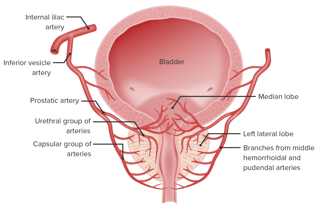
Where are the Leydig cells located?
- In the interstitial spaces, adjacent to the seminiferous tubules
- Inside the seminiferous tubules
- Adjacent to the seminal vesicles
- Inside the seminal vesicles
- Within the epididymis
Author of lecture Testis: Seminiferous Tubule – Male Reproductive System

Geoffrey Meyer, PhD
Customer reviews
5,0 of 5 stars
| 5 Stars |
|
5 |
| 4 Stars |
|
0 |
| 3 Stars |
|
0 |
| 2 Stars |
|
0 |
| 1 Star |
|
0 |
