Playlist
Show Playlist
Hide Playlist
Gluteal Region – Gluteal Region and Posterior Thigh


-
Slides 04 LowerLimbAnatomy Pickering.pdf
-
Download Lecture Overview
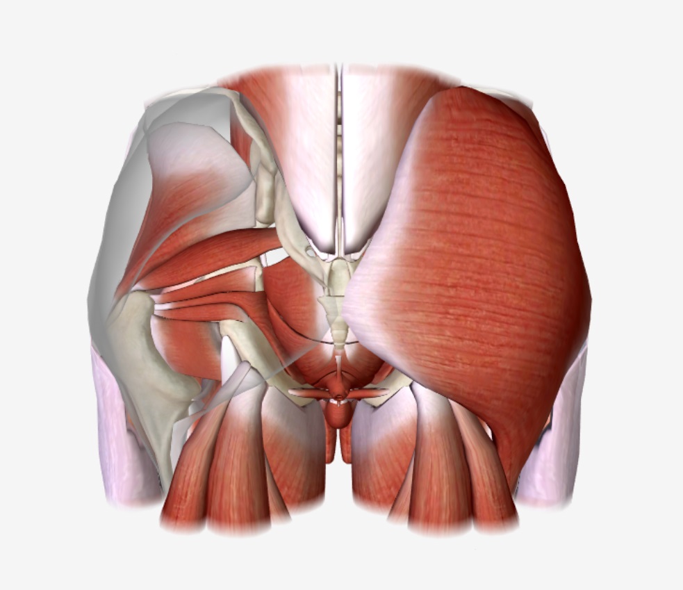
00:01 Within the posterior thigh, we have a whole series of muscles. We have gluteus maximus, we have gluteus medius, and we have gluteus minimus. I?m just going to concentrate on these, first of all. This is the posterior aspect of the right lower limb. So we can see we?ve got the iliac crest here, we?ve got the sacrum, and then we?ve got the posterior shaft of the femur. We can see gluteus maximus, and then we can see gluteus medius and gluteus minimus. 00:30 And these muscles are all passing inferolaterally towards the femur. Gluteus medius and gluteus minimus are running down onto the greater trochanter, whereas, gluteus maximus passes through the iliotibial tract, and also to the gluteal tuberosity. So here we can see we have gluteus maximus, we have gluteus medius, we have gluteus minimus, and we can see their origins and their insertions. We can see for gluteus maximus, it?s coming from the ilium posterior to the gluteal line. It?s also coming from the posterior sacrum and coccyx and sacrotuberous ligaments. We can see that here, it?s coming from the posterior ilium, posterior to that posterior line. It?s coming from the sacrum, and it?s also coming from parts of the coccyx and sacrotuberous ligament. It then passes inferolaterally, and we can see it is passing most of it to the iliotibial tract, about 75%. The deeper quarter, so the deeper muscle fibres are passing through the gluteal tuberosity. It?s innervated via the inferior gluteal nerve, and it?s important in extending the hip, and it also assists in lateral rotation. So it?s an important extender of the hip. Gluteus medius, we can see coming from the external surface of the ilium, this time between the anterior and posterior gluteal lines. So it?s important to remember these gluteal lines now. 02:02 We can see gluteus medius coming from between the anterior and the posterior gluteal lines, and it?s passing towards the greater trochanter of the femur. Gluteus minimus comes from, again, the external surface of the ilium between the anterior and inferior gluteal lines, and this also passes towards the greater trochanter of the femur. We can see the gluteus minimus coming again here now towards the greater tubercle. So we got gluteus maximus, gluteus medius, and gluteus minimus. Gluteus medius and gluteus minimus are innervated via the superior gluteal nerve, and they are involved in abducting and medially rotating the femur, abducting the femur with the hip joints and also medially rotating the hip joint. 02:55 They?re also involved in keeping the pelvis level when the opposite limb is off the ground. 03:01 This is important to prevent tilting of the pelvis when you?re walking. So we?ll see this as we go through the course when we look at the nerve lesions. The final muscle I want to draw a reference to is the tensor fasciae latae, and this alongside gluteus maximus passes into the iliotibial tract. So we can see tensor fasciae latae here. Tensor fasciae latae is coming from the anterior superior iliac spine, and it passes to the iliotibial tract which goes to the lateral condyle of the tibia. It?s also innervated via the superior gluteal nerve. It is involved in flexing the hip and also stabilizing the knee joint. Now let?s look at the muscles that lie deep to the gluteal muscles, of at least gluteus maximus and gluteus medius. We?re going to look at piriformis, the gemelli, obturator internus, and quadratus femoris. Here, we can see we?ve removed gluteus medius and we can now reveal piriformis, this muscle passing out of the pelvis through the greater sciatic foramen. We can see we got piriformis. This is passing through the greater trochanter. 04:13 We can then see we have two gemelli muscles. We have superior gemellus and we have inferior gemellus. And here, we also have the tendon of obturator internus. So here we can see obturator internus, just the tendon really. This is the inside. This is the posterior view of the hip. This is the internal surface of obturator membrane. And this is actually sending a tendon away from that muscle belly and through the lesser sciatic foramen to the greater trochanter. We can see that this tendon is running in between the superior and inferior gemelli muscles. Most inferiorly, we find we have quadratus femoris, and that?s running from the ischial tuberosity across through the intertrochanteric crest on the posterior surface. We can see we have piriformis, the superior gemellus, obturator internus, inferior gemellus, and then quadratus femoris. What we can see is the origins and insertions in this table. We have piriformis coming from the anterior surface of the sacrum and passing to the greater trochanter. We have the two gemelli, superior coming from the ischial spine, and inferior coming from the ischial tuberosity. These also pass to the greater trochanter. Obturator internus, this is coming from the pelvic surface, as I said, of the ilium and the ischium, and the obturator membrane lining the obturator foramen. And this also is passing to the greater trochanter. These were all supplied by specific nerves. 05:52 These are the root values, and the nerves known as to that specific muscle. So nerve to obturator internus comes from S1. Nerve to quadratus femoris comes from L5, S1. And nerve to obturator internus comes also from S1. Quadratus femoris, this is coming from the ischial tuberosity and it passes, as I said, to the intertrochanteric crest. This again is coming to nerve supply from L5 to S1. All of these muscles are known as lateral rotators of the hip. So lateral rotators of the hip, especially when it?s extended. They?re also involved in abducting the hip, so drawing it away from the body. And this should be obvious from their attachment onto the greater trochanter. The quadratus femoris as well as being a lateral rotator, it also helps to hold the head of the femur in the acetabulum. So if we look at these in a bit more detail with lots of the muscles all in place, then we can see we?ve got the posterior surface of the gluteal region here, and a deeper dissection here. 07:03 We can see that we have a mass of muscles on the posterior gluteal region. When all of these muscles are put in place, a mass of musculature. We have gluteus maximus that has been removed, we have gluteus medius, and we have gluteus minimus. And then we have these lateral rotators and hip stabilizers. Most laterally, we can see tensor fasciae latae. If we?re then to see in more detail the fan-shaped gluteus medius, we can see here coming away from the ilium, and then deep to it, we have gluteus minimus. Gluteus maximus, this large muscle, has been removed. Laterally, we?ve got the tensor fasciae latae muscle, and this helps to stabilize the lateral aspect of the knee joint.
About the Lecture
The lecture Gluteal Region – Gluteal Region and Posterior Thigh by James Pickering, PhD is from the course Lower Limb Anatomy [Archive].
Included Quiz Questions
Which muscle passes through the greater sciatic foramen?
- Piriformis
- Obturator externus
- Obturator internus
- Superior gemellus
- Inferior gemellus
Into which anatomical location does the gluteus medius insert?
- Greater trochanter of femur
- Gluteal tuberosity
- Iliotibial tract
- Anterior iliac spine
- External ilium
Which location is the correct anatomical site of the origin of the quadratus femoris?
- Ischial tuberosity
- Ischial spine
- Greater trochanter
- Anterior surface of sacrum
- Obturator membrane
Author of lecture Gluteal Region – Gluteal Region and Posterior Thigh

James Pickering, PhD
Customer reviews
5,0 of 5 stars
| 5 Stars |
|
1 |
| 4 Stars |
|
0 |
| 3 Stars |
|
0 |
| 2 Stars |
|
0 |
| 1 Star |
|
0 |
I love the explanation. Very easy to understand and remember.
