Playlist
Show Playlist
Hide Playlist
Derma Case: 11-year-old Healthy Girl with Rash

-
Slides Virally-mediated Skin Lesions.pdf
-
Download Lecture Overview
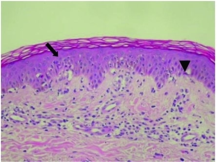
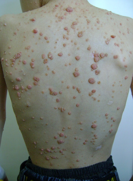
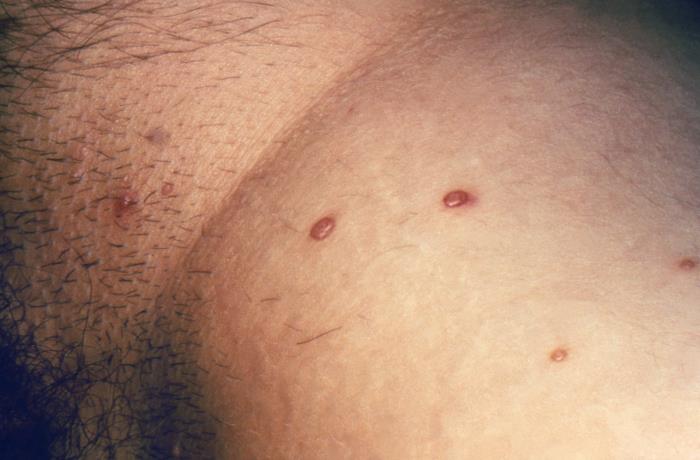
00:01 Alright, we're gonna cover a big topic today, virally mediated skin lesions. 00:07 Let's get started, as we always do, with a case. 00:10 An 11 year old healthy girl presents with a rash. 00:13 Her mother states that she's noticed a crop of round bumps appearing on her trunk and her thighs over the past several days and particularly in the groin and in her armpits. 00:23 The mother appears more distressed about them than the daughter, who reports that they aren't bothering her at all. 00:28 Patient denies any fevers, chills, myalgias, arthralgias, URI symptoms or GI symptoms. 00:35 The only thing is that some of the lesions are very mildly itchy. 00:39 Social and family history - non contributory. 00:42 Review of systems as we've discussed is totally negative and her vital signs are completely normal. 00:47 What we see on exam is numerous subcentimeter, skin colored, dome-shaped papules scattered on her torso and we can see some here on the picture on the right. 00:57 They're also in the axillae, the crural folds, the proximal thighs. 01:01 The lesions are very shiny in appearance, umbilicated, they're non-tender and they're firm, sparing the palms and the soles. 01:09 And importantly, there's no evidence of any mucosal lesions. 01:13 So, let's look at a few key points here, highlighting some prominent variables. 01:18 Number 1, they sound acute. 01:19 They've just been coming on over the past couple of days. 01:23 Secondly, the pattern of skin involvement, they're located on her trunk, in her thighs and rather asymmetrically distributed. 01:32 Skin inflammation, the fact that they're non-tender, there are not any postules. 01:37 It doesn't sound like there's any skin inflammation and there really is no evidence of any skin involvement or systemic involvement based on the comprehensive review of systems. 01:45 Alright, so let's take a look at our differential diagnosis for this case. 01:49 First stop, keratoacanthoma. 01:51 Now, keratoacanthomas are indeed often described as dome-shaped firm papules. 01:58 There's one right there and it does kinda look like what she has. 02:02 And they'ren asymptomatic which is also similar to what she has. 02:05 But they don't appear in crops as they did in our patient. 02:09 They are isolated, individual lesions with a central keratotic plug. 02:14 Moreover, you'd almost never see one in an 11-year-old girl. 02:17 It's a disease of folks over the age of 50. 02:20 It represents a type of very low grade malignancy, sometimes called a pseudocancer. 02:25 Now, in defense of it's appearance in this section on viral diseases of the skin, I would be remiss if I did not also mention that it is strongly associated with HPV. 02:36 So, it's a reasonable thing to have in this topic, but it's definitely not what she has, so let's X it out. 02:43 Next stop, varicella, also known as chickenpox. 02:47 Well, it's one of the many viruses that can cause a so-called viral exanthem, dozen of viruses can do this. 02:53 Now, the lesions of chickenpox can come on fairly quickly, just like our patient did, over several days. 02:58 And they might indeed look like round, dome-shaped lesions. 03:03 But the lesions of chicken pox are actually fluid-filled vesicles rather than these firm papules. 03:09 And varicella, like most viral exanthems is accompanied by symptoms: Fevers, malaise, myalgias, headaches, perhaps even a sore throat. 03:17 I had it when I was 19, it was terrible. 03:21 Now less commonly, viral exanthems could have diarrhea, lymphadenopathy, maybe even splenomegaly or hepatomegaly, Either way, it doesn't sound like what she's got. 03:31 Varicella lesions often transition from vesicles to pustules after a few days and ultimately will cross over and resolve spontaneously in less than 10 days. 03:39 Just rounding out the discussion about varicella. 03:42 So, I think we can safely take it off of our list for now. 03:47 Molluscum contagiosum. 03:49 Now, this rash which is also caused by a virus, the molluscum contagiosum virus is relatively common in children. 03:58 The lesions of molluscum contagiosum are described as dome-shaped papules, randomly distributed on the torso and extremities in an asymptomatic host. 04:08 Looks like we'll need to keep this one on the list for now. 04:11 Those lesions do look familiar. 04:15 Next stop, measles. 04:17 Okay, this is yet another virus that causes a viral exanthem and like varicella, it's typically accompanied by a viral prodrome. 04:25 Fevers, malaise, cough, conjunctivitis is common and sinus congestion. 04:30 Now, the lesions themselves are typically diffuse, erythematous maculopapules, often referred to as 'morbilliform' because many other viruses look the same as measles. 04:41 They're only a few millimeters in size, shown here in the top right picture and they typically start on the face, spreading caudally with areas of confluence often on the chest and the neck. 04:51 Shown here on the bottom right is a reminder that measles is also associated with classic mucosal lesions called 'Koplik spots', you'll definitely see this referenced on the boards. 05:01 They are grayish, white, flat macules on an erythematous base, seen on the buccal mucosa adjacent to the molars as shown here. 05:10 Now, our patient had firm, dome-shaped papules. 05:14 No mucosal lesions, not to mention she was completely asymptomatic. 05:17 So I think we can definitely take this one off of our list. 05:22 Lastly, let's talk about erythema multiforme. 05:25 Remember, while we often think of erythema multiforme along with Steven-Johnsons Syndrome and toxic epidermal necrolysis associated with drug reactions, keep in mind that 90% of erythema multiforme eruptions are actually due to infections, most notably HSV and then mycoplasma would be second in place. 05:44 Let's take a look at the next slide to review erythema multiforme in some more detail. 05:49 So, here's some picture shown in the right of classic erythema multiforme manifestations. 05:55 It is an acute, immune-mediated eruption with targetoid-shaped lesions depicted here. 06:01 It's really gonna afflict young adults, predominantly under age 30, oftentimes, under age 10. 06:07 90% of them as I mentioned are caused by infections like HSV and mycoplasma. 06:12 Less than 10% of the time, it's caused by drugs. 06:15 As we'll see in some other lecture talking about SJS and toxic epidermal necrolysis that ratio is flipped with a small number causing, or caused by infections and a much larger number caused by medications. 06:28 What you'll see here on the right are these targetoid erythematous papules that are symmetrically distributedon the hands and the face with a centripetal spread towards the torso. 06:38 And you can note here the central clearing on some of those lesions. 06:41 As a quick aside, sometimes I have trouble remembering what centripetal versus cetrifugal means. 06:46 Always remember cetrifugal has F-U-G, and that makes me think of a fugitive. 06:52 What are fugitives doing? they're running away. 06:54 So centrifugal means striding away from the body as opposed to centripetal is running towards the body. 07:00 or coming towards the body. 07:02 Important for us to remember that there are two subtypes of erythema multiforme: the minor and the major subtype. 07:08 The minor subtype is basically characterized by just the rash alone, whereas the major subtype is more like a Steven-Johnsons syndrome type of picture with mucosal involvement, malaise, myalgias, fevers, other constitutional symptoms and it's much more significant of course. 07:24 Our patient, clearly, if anything would be looking like a minor variant here. 07:30 The way that it's managed, if it's mediatied or caused by herpes simplex virus, you're gonna want to treat that. 07:35 Withdraw whatever culprit medications could be playing a role and you may need to add on systemic glucocorticoids, generally just for the major subtype of erythema multiforme. 07:44 So, having reviewed erythema multiforme, I think we can safely take it off our list as the lesions are wholly inconsistent with our patient's dome-shaped, skin-colored papules. 07:57 Alright, so it looks like we're left with molluscum cotagiosum. 08:00 So let's revisit the case and highlight some key points. 08:05 Alright, so molluscum contagiosum is very common in children and the immunocompromised and our 11-year-old healthy girl would be a perfect case of somebody getting molluscum contagiosum. 08:17 The lesions here, the round bumps appearing on her trunk and thighs over the past several days particularly the groin and the armpits, this is a classic presentation as well. 08:26 Molluscum contagiosum lesions are asymmetric, they've predilection for flexural creases and they spare the palms, the soles and there should not be any mucosal involvement. 08:37 Next stop, as we've highlighted countless times over the past few minutes, patients should be asymptomatic. 08:43 They shouldn't be manifesting with the classic viral syndrome of myalgias and fevers that sort of thing. 08:50 Next stop, the lesions as we've said are dome-shaped papules with these characterized central umblication, a little bit of a hallowing out in the center of the lesion and they're non-tender.
About the Lecture
The lecture Derma Case: 11-year-old Healthy Girl with Rash by Stephen Holt, MD, MS is from the course Skin Infections.
Included Quiz Questions
Which of the following best describes varicella lesions?
- Fluid-filled vesicular lesions that transform into pustules
- Dome-shaped papules with central umbilication
- Itchy, erythematous papules
- Maculopapular lesions with silvery scales
- Blood-filled vesicles and bullae
Which of the following is true regarding measles?
- It is characterized by white macules on an erythematous base may be seen on the buccal mucosa.
- It is accompanied by distal muscle weakness.
- It is caused by herpes simplex virus 1.
- It presents as cutaneous, fluid-filled vesicles.
- Skin lesions usually start on soles and palms that then spread centrally.
Erythema multiforme is commonly associated with which of the following viruses?
- Herpes simplex virus
- Measles virus
- Human immunodeficiency virus
- Human papillomavirus
- Hepatitis B virus
A 22-year-old Caucasian man presents to the clinic with multiple erythematous macules and papules. Moreover, the skin lesions were first noticed over his hands and face and then spread toward his trunk. On physical examination, they appear as targetoid skin lesions. His medical history is significant for mycoplasma pneumonia infection 1 week ago. Which of the following is the most likely diagnosis?
- Erythema multiforme
- Psoriasis
- Pemphigus vulgaris
- Urticaria
- Erythema migrans
Which of the following statements is true regarding molluscum contagiosum?
- It is caused by the molluscum contagiosum virus.
- It mainly affects the elderly.
- Fever and arthralgias usually precede cutaneous eruptions.
- Symmetrical distribution all over the body is the rule.
- Palms and soles are usually affected.
Author of lecture Derma Case: 11-year-old Healthy Girl with Rash

Stephen Holt, MD, MS
Customer reviews
5,0 of 5 stars
| 5 Stars |
|
1 |
| 4 Stars |
|
0 |
| 3 Stars |
|
0 |
| 2 Stars |
|
0 |
| 1 Star |
|
0 |
Very informative, I found the photos and discussion of differentials very helpful.
