Playlist
Show Playlist
Hide Playlist
Foot – Osteology of Lower Limb

-
Slides 01 LowerLimbAnatomy Pickering.pdf
-
Download Lecture Overview
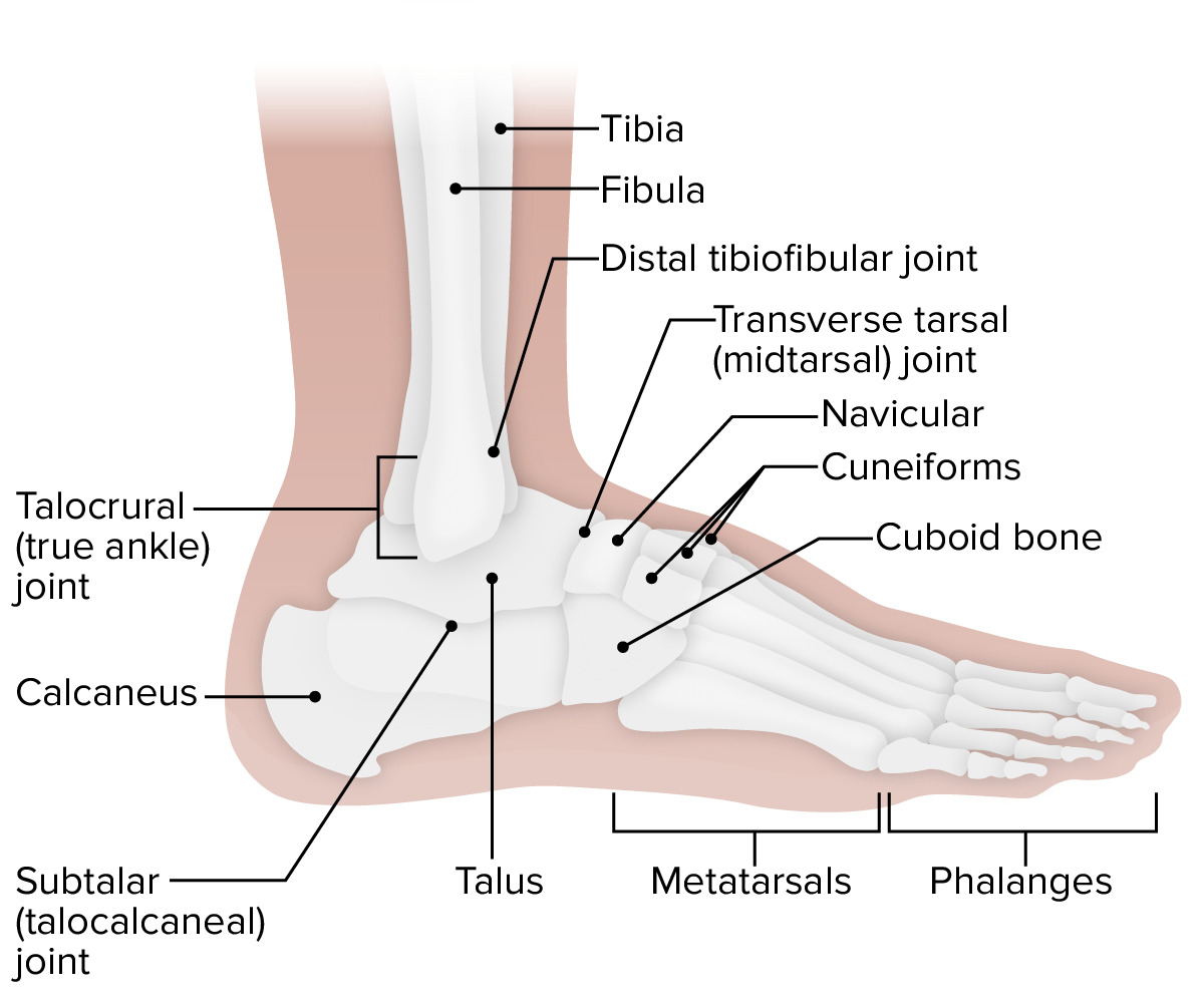
00:01 Now let’s move on to the foot and look at the numerous bones that make up the foot, both the tarsals, the metatarsals, and the phalanges. 00:11 We’ve got a whole series of pictures that show different views of the foot. We can see that we have tarsus, these seven bones that help to form the main body of the foot, and then we can see we have the metatarsus that contains these metatarsals, and then we can see we have the phalanges. 00:32 But for the tarsus, we can have the calcaneus, the talus, navicular, cuboid, and three cuneiforms. 00:40 Here, we can locate, if we look more superiorly, the talus, and this is going to allow articulation between the tibia and the fibula via the two malleoli. Then inferior to the talus underneath, we have the calcaneus. We can see the calcaneus is then going to articulate with the cuboid, we can see here. And directly in front of the talus, we can see we have the navicular. 01:07 So we’ve got the talus here. We’ve got the navicular. And then we move lateral and we find the cuboid. We then have three cuneiforms that are positioned anterior to the navicular bone. 01:20 We have the medial, the intermediate, and the lateral. So we have the talus, we have the calcaneus here, we have the navicular, the cuboid, and then three cuneiforms - medial, intermediate, and lateral. If we then look at the metatarsus, here we have five metatarsals. 01:41 These connect to the phalanges, and toe 1 is the shortest metatarsus. So it’s not as long as the 2, 3, 4, and 5. So the phalanges had a similar arrangement as we had in the hand. So toe 1 has two phalanges, a proximal and a distal. And toe 2 to 5 has a proximal, a middle, and a distal. And this is repeated from toes 2, 3, 4, and 5. 02:14 So let’s have a look at a bit more detail at these tarsal bones. Let’s start off with the calcaneus. We can see it’s the largest, the strongest of the tarsal bone, and articulates with the talus superiorly and the cuboid bone anteriorly. We can see it here. We can see if we have a plantar view here, a lateral view and a dorsal view. This is the calcaneus. 02:41 Medially, if we look at this medial aspect of the calcaneus, we see it forms what’s known as the sustentaculum tali. And this helps to support the talus. It’s like a shelf and it helps to support the talus superiorly, the sustentaculum tali. We’ll later on appreciate some important tendons, blood vessels and nerves pass underneath the sustentaculum tali as they cross posteriorly to the medial malleolus to the enter the sole of the foot. 03:12 We also have the calcaneal tuberosity. We can see that here, this large inferior bulge, the calcaneal tuberosity, and that’s an important attachment site for the calcaneal tendon. 03:26 So now if we look at the talus, then that sits superior to the calcaneus, and we have a head, we have a neck, and we have a body. So we can see the neck of the talus here in this lateral view, and if we move on to the medial view, we see most anteriorly, we have the head, then we have the neck, and then we can see we have posteriorly the body. 03:52 We can see how it’s passing down and attaching to the calcaneus in line with the sustentaculum tali. 03:58 And here, we can see the talus. The trochlear surface which we can see here, so we have the trochlear surface similar to the trochlear that we have in the upper limb, allows for articulation with the two malleoli coming from the tibia and the fibula. 04:16 And we’ll appreciate that this is wedge shaped allowing for strengthening of the ankle joint during various movements. Now let’s turn to the navicular. This is a flattened boat shaped bone. And it is located between the talus posteriorly, and the three cuneiforms anteriorly. We can see it here clearly in this dorsal view. On the lateral view, we can see it’s lying medial to the cubiod bone, and on this medial view, we can see it’s running against the medial surface of the foot. On the inferior surface of the navicular, we see a medially orientated tuberosity, and these were important for muscle attachments. 05:00 If we then look at the cuboid bone, we can see the cuboid is located in between the calcaneus, and the fourth and fifth metatarsals. Medially, we would have the lateral cuneiform bone. 05:15 So here we can see the cuboid. The cuboid itself has a tuberosity which we can again see on the plantar view, and we also see it has a groove for the fibularis longus tendon. 05:28 We’d appreciate that when we look at the fibula muscles in a later lecture. If we now move to the cuneiforms, now these forms a row of bones that is directly anterior to the navicular. And we have from medial to lateral, we have a medial, intermediate, and a lateral cuneiform. We can see the three here in this dorsal view, and we can see the three here in this plantar view. All of these three bones articulate with the navicular posteriorly and the metatarsals anteriorly. So now let’s talk about those metatarsals. We have five metatarsals, and these run forward to connect to the phalanges of the digits. Toe 1 has the shortest metatarsal, and we can see that here. It’s very large, it’s very substantial, it’s made up of a base, it’s made up of a shaft, and it’s made up of a head. 06:26 So we have this very broad large first metatarsal, but it’s also the shortest. If we look at metatarsals 2, 3, 4, and 5, we see they have a base, a shaft, and a head, but these are much thinner. We can then see just like the digits in the hand, digit 1 has two phalanges, a proximal and a distal. So we can see the proximal and distal phalanges of the first digit, the toe. And then digits 2, 3, 4, and 5 have both proximal, have all proximal, middle, and distal phalanges.
About the Lecture
The lecture Foot – Osteology of Lower Limb by James Pickering, PhD is from the course Lower Limb Anatomy [Archive].
Included Quiz Questions
Which bone is part of the tarsus?
- Calcaneus
- Phalanges
- Metatarsus
- Lunate
- Hamate
Which bone is the most anatomically posterior?
- Calcaneus
- Talus
- Navicular
- Cuboid
- Medial cuneiform
Author of lecture Foot – Osteology of Lower Limb

James Pickering, PhD
Customer reviews
5,0 of 5 stars
| 5 Stars |
|
3 |
| 4 Stars |
|
0 |
| 3 Stars |
|
0 |
| 2 Stars |
|
0 |
| 1 Star |
|
0 |
superb lectures with transcript, 3d models and as many study resources.
It’s very good I really enjoyed this lecture.the study of way is very good
video was simple and fast, and well articulated. Much appreciated
